 Movie
Movie Controller


+ Open data

- Basic information
| Entry | Database: PDB / ID: 8f0n | ||||||
|---|---|---|---|---|---|---|---|
| Title | Wobble Beetroot (A16U-U38G) dimer bound to DFHO | ||||||
 Components Components | RNA (49-MER) | ||||||
 Keywords Keywords | RNA / aptamer / fluorescence / turn-on / fluorogenic / fluorophore / G-quartet / G-quadruplex | ||||||
| Function / homology | Chem-747 / : / RNA / RNA (> 10) Function and homology information Function and homology information | ||||||
| Biological species | synthetic construct (others) | ||||||
| Method |  X-RAY DIFFRACTION / SYNCHROTRON / MOLECULAR REPLACEMENT / Resolution: 2.85 Å X-RAY DIFFRACTION / SYNCHROTRON / MOLECULAR REPLACEMENT / Resolution: 2.85 Å | ||||||
 Authors Authors | Passalacqua, L.F.M. / Ferre-D'Amare, A.R. | ||||||
| Funding support |  United States, 1items United States, 1items
| ||||||
 Citation Citation | Journal: Nat Commun / Year: 2023 Title: Co-crystal structures of the fluorogenic aptamer Beetroot show that close homology may not predict similar RNA architecture. Authors: Passalacqua, L.F.M. / Starich, M.R. / Link, K.A. / Wu, J. / Knutson, J.R. / Tjandra, N. / Jaffrey, S.R. / Ferre-D'Amare, A.R. | ||||||
| History |
|
- Structure visualization
| Structure viewer | Molecule: MolmilJmol/JSmol |
|---|
- Downloads & links
-Download
| PDBx/mmCIF format | 8f0n.cif.gz | 143 KB | Display | PDBx/mmCIF format |
|---|---|---|---|---|
| PDB format | pdb8f0n.ent.gz | 94.1 KB | Display | PDB format |
| PDBx/mmJSON format | 8f0n.json.gz | Tree view | PDBx/mmJSON format | |
| Others |  Other downloads Other downloads |
-Validation report
| Arichive directory | https://data.pdbj.org/pub/pdb/validation_reports/f0/8f0nftp://data.pdbj.org/pub/pdb/validation_reports/f0/8f0n | HTTPS FTP |
|---|
-Related structure data
| Related structure data | 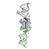 8eyuSC 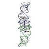 8eyvC 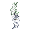 8eywC S: Starting model for refinement C: citing same article ( |
|---|---|
| Similar structure data |
-Links
 PDBj
PDBj


- Assembly
| Deposited unit | 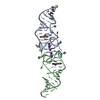
| ||||||||||||
|---|---|---|---|---|---|---|---|---|---|---|---|---|---|
| 1 |
| ||||||||||||
| Unit cell |
|
-Components
| #1: RNA chain | Mass: 16039.509 Da / Num. of mol.: 2 / Mutation: A16U, U38G / Source method: obtained synthetically / Source: (synth.) synthetic construct (others) #2: Chemical | ChemComp-K /   Mass: 39.098 Da / Num. of mol.: 5 / Source method: obtained synthetically / Formula: K Mass: 39.098 Da / Num. of mol.: 5 / Source method: obtained synthetically / Formula: K#3: Chemical |   Mass: 281.215 Da / Num. of mol.: 2 / Source method: obtained synthetically / Formula: C12H9F2N3O3 / Feature type: SUBJECT OF INVESTIGATION Mass: 281.215 Da / Num. of mol.: 2 / Source method: obtained synthetically / Formula: C12H9F2N3O3 / Feature type: SUBJECT OF INVESTIGATION#4: Water | ChemComp-HOH / |  Mass: 18.015 Da / Num. of mol.: 35 / Source method: isolated from a natural source / Formula: H2O Mass: 18.015 Da / Num. of mol.: 35 / Source method: isolated from a natural source / Formula: H2OHas ligand of interest | Y | |
|---|
-Experimental details
-Experiment
| Experiment | Method: X-RAY DIFFRACTION / Number of used crystals: 1 |
|---|
- Sample preparation
| Crystal | Density Matthews: 2.21 Å3/Da / Density % sol: 44.23 % |
|---|---|
| Crystal grow | Temperature: 294 K / Method: vapor diffusion, hanging drop / pH: 7 / Details: 2.4 M Ammonium sulfate, 5% 2-propanol |
-Data collection
| Diffraction | Mean temperature: 100 K / Serial crystal experiment: N |
|---|---|
| Diffraction source | Source: SYNCHROTRON / Site: ALS / Beamline: 5.0.2 / Wavelength: 1 Å |
| Detector | Type: DECTRIS PILATUS3 6M / Detector: PIXEL / Date: Dec 10, 2021 |
| Radiation | Protocol: SINGLE WAVELENGTH / Monochromatic (M) / Laue (L): M / Scattering type: x-ray |
| Radiation wavelength | Wavelength: 1 Å / Relative weight: 1 |
| Reflection | Resolution: 2.85→94.12 Å / Num. obs: 6710 / % possible obs: 99.88 % / Redundancy: 6.4 % / Biso Wilson estimate: 42.69 Å2 / CC1/2: 0.989 / Rmerge(I) obs: 0.295 / Rpim(I) all: 0.126 / Rrim(I) all: 0.321 / Net I/σ(I): 2.5 |
| Reflection shell | Resolution: 2.85→2.95 Å / Redundancy: 5.9 % / Rmerge(I) obs: 1.186 / Num. unique obs: 617 / CC1/2: 0.482 / % possible all: 90.47 |
- Processing
| Software |
| ||||||||||||||||||||||||||||||||||||||||||
|---|---|---|---|---|---|---|---|---|---|---|---|---|---|---|---|---|---|---|---|---|---|---|---|---|---|---|---|---|---|---|---|---|---|---|---|---|---|---|---|---|---|---|---|
| Refinement | Method to determine structure: MOLECULAR REPLACEMENT Starting model: 8EYU Resolution: 2.85→47.06 Å / SU ML: 0.4284 / Cross valid method: FREE R-VALUE / σ(F): 1.33 / Phase error: 23.1153 Stereochemistry target values: GeoStd + Monomer Library + CDL v1.2
| ||||||||||||||||||||||||||||||||||||||||||
| Solvent computation | Shrinkage radii: 0.9 Å / VDW probe radii: 1.11 Å / Solvent model: FLAT BULK SOLVENT MODEL | ||||||||||||||||||||||||||||||||||||||||||
| Displacement parameters | Biso mean: 59.48 Å2 | ||||||||||||||||||||||||||||||||||||||||||
| Refinement step | Cycle: LAST / Resolution: 2.85→47.06 Å
| ||||||||||||||||||||||||||||||||||||||||||
| Refine LS restraints |
| ||||||||||||||||||||||||||||||||||||||||||
| LS refinement shell |
| ||||||||||||||||||||||||||||||||||||||||||
| Refinement TLS params. | Method: refined / Origin x: 5.72681320393 Å / Origin y: -7.68783291633 Å / Origin z: 3.92979116507 Å
| ||||||||||||||||||||||||||||||||||||||||||
| Refinement TLS group | Selection details: all |
