 Movie
Movie Controller

[English] 日本語
 Yorodumi
- PDB-8ewy: Structure of Janus Kinase (JAK) dimer complexed with cytokine rec... -
+ Open data

- Basic information
| Entry | Database: PDB / ID: 8ewy | ||||||
|---|---|---|---|---|---|---|---|
| Title | Structure of Janus Kinase (JAK) dimer complexed with cytokine receptor intracellular domain | ||||||
 Components Components |
| ||||||
 Keywords Keywords | SIGNALING PROTEIN / JAK / Kinase / Cytokine | ||||||
| Function / homology |  Function and homology information Function and homology informationresponse to type III interferon / interleukin-28 receptor complex / Interleukin-20 family signaling / mucosal immune response / positive regulation of cellular respiration / protein localization to cell-cell junction / interleukin-11-mediated signaling pathway / CCR5 chemokine receptor binding / interleukin-9-mediated signaling pathway / interleukin-4-mediated signaling pathway ...response to type III interferon / interleukin-28 receptor complex / Interleukin-20 family signaling / mucosal immune response / positive regulation of cellular respiration / protein localization to cell-cell junction / interleukin-11-mediated signaling pathway / CCR5 chemokine receptor binding / interleukin-9-mediated signaling pathway / interleukin-4-mediated signaling pathway / interleukin-10-mediated signaling pathway / interleukin-2-mediated signaling pathway / positive regulation of homotypic cell-cell adhesion / regulation of defense response to virus by host / interleukin-15-mediated signaling pathway / cytokine receptor activity / growth hormone receptor binding / extrinsic component of cytoplasmic side of plasma membrane / type I interferon-mediated signaling pathway / interleukin-6-mediated signaling pathway / positive regulation of sprouting angiogenesis / cell surface receptor signaling pathway via JAK-STAT / type II interferon-mediated signaling pathway / non-membrane spanning protein tyrosine kinase activity / non-specific protein-tyrosine kinase / positive regulation of protein localization to nucleus / protein phosphatase binding / defense response to virus / cytoskeleton / receptor complex / intracellular signal transduction / response to antibiotic / ubiquitin protein ligase binding / ATP binding / nucleus / cytoplasm Similarity search - Function | ||||||
| Biological species |  | ||||||
| Method | ELECTRON MICROSCOPY / single particle reconstruction / cryo EM / Resolution: 5.5 Å | ||||||
 Authors Authors | Caveney, N.A. / Saxton, R.A. / Waghray, D. / Garcia, K.C. | ||||||
| Funding support |  United States, 1items United States, 1items
| ||||||
 Citation Citation | Journal: Cell Rep / Year: 2023 Title: Structural basis of Janus kinase trans-activation. Authors: Nathanael A Caveney / Robert A Saxton / Deepa Waghray / Caleb R Glassman / Naotaka Tsutsumi / Stevan R Hubbard / K Christopher Garcia / Abstract: Janus kinases (JAKs) mediate signal transduction downstream of cytokine receptors. Cytokine-dependent dimerization is conveyed across the cell membrane to drive JAK dimerization, trans- ...Janus kinases (JAKs) mediate signal transduction downstream of cytokine receptors. Cytokine-dependent dimerization is conveyed across the cell membrane to drive JAK dimerization, trans-phosphorylation, and activation. Activated JAKs in turn phosphorylate receptor intracellular domains (ICDs), resulting in the recruitment, phosphorylation, and activation of signal transducer and activator of transcription (STAT)-family transcription factors. The structural arrangement of a JAK1 dimer complex with IFNλR1 ICD was recently elucidated while bound by stabilizing nanobodies. While this revealed insights into the dimerization-dependent activation of JAKs and the role of oncogenic mutations in this process, the tyrosine kinase (TK) domains were separated by a distance not compatible with the trans-phosphorylation events between the TK domains. Here, we report the cryoelectron microscopy structure of a mouse JAK1 complex in a putative trans-activation state and expand these insights to other physiologically relevant JAK complexes, providing mechanistic insight into the crucial trans-activation step of JAK signaling and allosteric mechanisms of JAK inhibition. | ||||||
| History |
|
- Structure visualization
| Structure viewer | Molecule: MolmilJmol/JSmol |
|---|
- Downloads & links
-Download
| PDBx/mmCIF format | 8ewy.cif.gz | 321.6 KB | Display | PDBx/mmCIF format |
|---|---|---|---|---|
| PDB format | pdb8ewy.ent.gz | 217.3 KB | Display | PDB format |
| PDBx/mmJSON format | 8ewy.json.gz | Tree view | PDBx/mmJSON format | |
| Others |  Other downloads Other downloads |
-Validation report
| Summary document | 8ewy_validation.pdf.gz | 1.3 MB | Display | wwPDB validaton report |
|---|---|---|---|---|
| Full document | 8ewy_full_validation.pdf.gz | 1.3 MB | Display | |
| Data in XML | 8ewy_validation.xml.gz | 52.6 KB | Display | |
| Data in CIF | 8ewy_validation.cif.gz | 84 KB | Display | |
| Arichive directory | https://data.pdbj.org/pub/pdb/validation_reports/ew/8ewyftp://data.pdbj.org/pub/pdb/validation_reports/ew/8ewy | HTTPS FTP |
-Related structure data
| Related structure data |  28649MC M: map data used to model this data C: citing same article ( |
|---|---|
| Similar structure data |
-Links
 PDBj
PDBj


- Assembly
| Deposited unit | 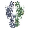
|
|---|---|
| 1 |
|
-Components
| #1: Protein | Mass: 136026.844 Da / Num. of mol.: 2 Source method: isolated from a genetically manipulated source Source: (gene. exp.)  Trichoplusia ni (cabbage looper) Trichoplusia ni (cabbage looper)References: UniProt: B1ASP2, non-specific protein-tyrosine kinase #2: Protein | Mass: 9895.373 Da / Num. of mol.: 2 Source method: isolated from a genetically manipulated source Source: (gene. exp.) Trichoplusia ni (cabbage looper) / References: UniProt: Q8CGK5#3: Chemical |   Mass: 267.241 Da / Num. of mol.: 2 / Source method: obtained synthetically / Formula: C10H13N5O4 Mass: 267.241 Da / Num. of mol.: 2 / Source method: obtained synthetically / Formula: C10H13N5O4#4: Chemical |   Mass: 427.201 Da / Num. of mol.: 2 / Source method: obtained synthetically / Formula: C10H15N5O10P2 / Comment: ADP, energy-carrying molecule*YM Mass: 427.201 Da / Num. of mol.: 2 / Source method: obtained synthetically / Formula: C10H15N5O10P2 / Comment: ADP, energy-carrying molecule*YMHas ligand of interest | N | Has protein modification | N | |
|---|
-Experimental details
-Experiment
| Experiment | Method: ELECTRON MICROSCOPY |
|---|---|
| EM experiment | Aggregation state: PARTICLE / 3D reconstruction method: single particle reconstruction |
- Sample preparation
| Component | Name: Structure of Janus Kinase (JAK) dimer complexed with cytokine receptor intracellular domain Type: COMPLEX / Entity ID: #1-#2 / Source: RECOMBINANT |
|---|---|
| Molecular weight | Experimental value: NO |
| Source (natural) | Organism: |
| Source (recombinant) | Organism: Trichoplusia ni (cabbage looper) |
| Buffer solution | pH: 8 |
| Specimen | Embedding applied: NO / Shadowing applied: NO / Staining applied: NO / Vitrification applied: YES |
| Vitrification | Cryogen name: ETHANE |
- Electron microscopy imaging
| Experimental equipment |  Model: Titan Krios / Image courtesy: FEI Company |
|---|---|
| Microscopy | Model: TFS KRIOS |
| Electron gun | Electron source:  FIELD EMISSION GUN / Accelerating voltage: 300 kV / Illumination mode: FLOOD BEAM FIELD EMISSION GUN / Accelerating voltage: 300 kV / Illumination mode: FLOOD BEAM |
| Electron lens | Mode: OTHER / Nominal defocus max: 3000 nm / Nominal defocus min: 800 nm |
| Image recording | Electron dose: 50.6 e/Å2 / Film or detector model: GATAN K3 BIOQUANTUM (6k x 4k) |
- Processing
| CTF correction | Type: PHASE FLIPPING AND AMPLITUDE CORRECTION |
|---|---|
| 3D reconstruction | Resolution: 5.5 Å / Resolution method: FSC 0.143 CUT-OFF / Num. of particles: 174962 / Symmetry type: POINT |
