 Movie
Movie Controller


+ Open data

- Basic information
| Entry | Database: PDB / ID: 8eww | ||||||
|---|---|---|---|---|---|---|---|
| Title | Structure of Arabidopsis fatty acid amide hydrolase mutant S305A | ||||||
 Components Components | Fatty acid amide hydrolase | ||||||
 Keywords Keywords | HYDROLASE / Fatty acid amide hydrolase / mutant / Arabidopsis / lipid signaling | ||||||
| Function / homology |  Function and homology information Function and homology informationN-(long-chain-acyl)ethanolamine deacylase activity / N-acylethanolamine metabolic process / fatty acid amide hydrolase / fatty acid amide hydrolase activity / plant-type vacuole / plastid / lipid catabolic process / defense response to bacterium / endoplasmic reticulum membrane / endoplasmic reticulum ...N-(long-chain-acyl)ethanolamine deacylase activity / N-acylethanolamine metabolic process / fatty acid amide hydrolase / fatty acid amide hydrolase activity / plant-type vacuole / plastid / lipid catabolic process / defense response to bacterium / endoplasmic reticulum membrane / endoplasmic reticulum / Golgi apparatus / membrane / plasma membrane Similarity search - Function | ||||||
| Biological species |  | ||||||
| Method |  X-RAY DIFFRACTION / SYNCHROTRON / MOLECULAR REPLACEMENT / Resolution: 2.8 Å X-RAY DIFFRACTION / SYNCHROTRON / MOLECULAR REPLACEMENT / Resolution: 2.8 Å | ||||||
 Authors Authors | Aziz, M. / Wang, X. / Gaguancela, O.A. / Chapman, K.D. | ||||||
| Funding support |  United States, 1items United States, 1items
| ||||||
 Citation Citation | Journal: To be published Title: Structural interactions explain the versatility of FAAH in the hydrolysis of plant and microbial acyl amide signals Authors: Aziz, M. / Wang, X. / Gaguancela, O.A. / Chapman, K.D. | ||||||
| History |
|
- Structure visualization
| Structure viewer | Molecule: MolmilJmol/JSmol |
|---|
- Downloads & links
-Download
| PDBx/mmCIF format | 8eww.cif.gz | 466.2 KB | Display | PDBx/mmCIF format |
|---|---|---|---|---|
| PDB format | pdb8eww.ent.gz | 390.7 KB | Display | PDB format |
| PDBx/mmJSON format | 8eww.json.gz | Tree view | PDBx/mmJSON format | |
| Others |  Other downloads Other downloads |
-Validation report
| Arichive directory | https://data.pdbj.org/pub/pdb/validation_reports/ew/8ewwftp://data.pdbj.org/pub/pdb/validation_reports/ew/8eww | HTTPS FTP |
|---|
-Related structure data
| Related structure data | 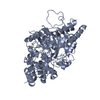 8ey1C 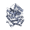 8ey9C 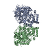 6dhvS S: Starting model for refinement C: citing same article ( |
|---|---|
| Similar structure data |
-Links
 PDBj
PDBj- Assembly
| Deposited unit | 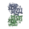
| ||||||||
|---|---|---|---|---|---|---|---|---|---|
| 1 | 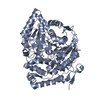
| ||||||||
| 2 | 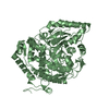
| ||||||||
| Unit cell |
|
-Components
| #1: Protein | Mass: 69563.141 Da / Num. of mol.: 2 / Mutation: S305A Source method: isolated from a genetically manipulated source Source: (gene. exp.)  #2: Water | ChemComp-HOH / |  Mass: 18.015 Da / Num. of mol.: 133 / Source method: isolated from a natural source / Formula: H2O Mass: 18.015 Da / Num. of mol.: 133 / Source method: isolated from a natural source / Formula: H2OHas protein modification | N | |
|---|
-Experimental details
-Experiment
| Experiment | Method: X-RAY DIFFRACTION / Number of used crystals: 1 |
|---|
- Sample preparation
| Crystal | Density Matthews: 2.5 Å3/Da / Density % sol: 50 % |
|---|---|
| Crystal grow | Temperature: 293 K / Method: vapor diffusion, hanging drop / pH: 6 / Details: 0.1 M MES, PH 6.0, 30% PEG 200, 5% PEG 3350 |
-Data collection
| Diffraction | Mean temperature: 93 K / Serial crystal experiment: N |
|---|---|
| Diffraction source | Source: SYNCHROTRON / Site: SSRL / Beamline: BL14-1 / Wavelength: 1.07809 Å |
| Detector | Type: MARMOSAIC 325 mm CCD / Detector: CCD / Date: Jul 23, 2019 |
| Radiation | Protocol: SINGLE WAVELENGTH / Monochromatic (M) / Laue (L): M / Scattering type: x-ray |
| Radiation wavelength | Wavelength: 1.07809 Å / Relative weight: 1 |
| Reflection | Resolution: 2.8→38.8 Å / Num. obs: 36725 / % possible obs: 98.8 % / Observed criterion σ(I): 1.4 / Redundancy: 3.5 % / CC1/2: 0.96 / Rmerge(I) obs: 0.044 / Net I/σ(I): 9.1 |
| Reflection shell | Resolution: 2.8→2.92 Å / Redundancy: 3.5 % / Rmerge(I) obs: 0.59 / Mean I/σ(I) obs: 1.4 / Num. unique obs: 4438 / CC1/2: 0.725 / % possible all: 98.1 |
- Processing
| Software |
| |||||||||||||||||||||||||||||||||||||||||||||||||||||||||||||||||||||||||||||||||||||||||||||||||||||||||||||||||||||||||||||||||||||||||||||||||||||||||||||||||||||||||||||||||||||||||||||||||||||||||||||||||||||||||||||||||
|---|---|---|---|---|---|---|---|---|---|---|---|---|---|---|---|---|---|---|---|---|---|---|---|---|---|---|---|---|---|---|---|---|---|---|---|---|---|---|---|---|---|---|---|---|---|---|---|---|---|---|---|---|---|---|---|---|---|---|---|---|---|---|---|---|---|---|---|---|---|---|---|---|---|---|---|---|---|---|---|---|---|---|---|---|---|---|---|---|---|---|---|---|---|---|---|---|---|---|---|---|---|---|---|---|---|---|---|---|---|---|---|---|---|---|---|---|---|---|---|---|---|---|---|---|---|---|---|---|---|---|---|---|---|---|---|---|---|---|---|---|---|---|---|---|---|---|---|---|---|---|---|---|---|---|---|---|---|---|---|---|---|---|---|---|---|---|---|---|---|---|---|---|---|---|---|---|---|---|---|---|---|---|---|---|---|---|---|---|---|---|---|---|---|---|---|---|---|---|---|---|---|---|---|---|---|---|---|---|---|---|---|---|---|---|---|---|---|---|---|---|---|---|---|---|---|---|
| Refinement | Method to determine structure: MOLECULAR REPLACEMENT Starting model: 6DHV Resolution: 2.8→38.78 Å / SU ML: 0.41 / Cross valid method: THROUGHOUT / σ(F): 1.35 / Phase error: 30.41 / Stereochemistry target values: ML
| |||||||||||||||||||||||||||||||||||||||||||||||||||||||||||||||||||||||||||||||||||||||||||||||||||||||||||||||||||||||||||||||||||||||||||||||||||||||||||||||||||||||||||||||||||||||||||||||||||||||||||||||||||||||||||||||||
| Solvent computation | Shrinkage radii: 0.9 Å / VDW probe radii: 1.11 Å / Solvent model: FLAT BULK SOLVENT MODEL | |||||||||||||||||||||||||||||||||||||||||||||||||||||||||||||||||||||||||||||||||||||||||||||||||||||||||||||||||||||||||||||||||||||||||||||||||||||||||||||||||||||||||||||||||||||||||||||||||||||||||||||||||||||||||||||||||
| Displacement parameters | Biso max: 177.41 Å2 / Biso mean: 99.4568 Å2 / Biso min: 41.23 Å2 | |||||||||||||||||||||||||||||||||||||||||||||||||||||||||||||||||||||||||||||||||||||||||||||||||||||||||||||||||||||||||||||||||||||||||||||||||||||||||||||||||||||||||||||||||||||||||||||||||||||||||||||||||||||||||||||||||
| Refinement step | Cycle: final / Resolution: 2.8→38.78 Å
| |||||||||||||||||||||||||||||||||||||||||||||||||||||||||||||||||||||||||||||||||||||||||||||||||||||||||||||||||||||||||||||||||||||||||||||||||||||||||||||||||||||||||||||||||||||||||||||||||||||||||||||||||||||||||||||||||
| LS refinement shell | Refine-ID: X-RAY DIFFRACTION / Rfactor Rfree error: 0 / Total num. of bins used: 13
| |||||||||||||||||||||||||||||||||||||||||||||||||||||||||||||||||||||||||||||||||||||||||||||||||||||||||||||||||||||||||||||||||||||||||||||||||||||||||||||||||||||||||||||||||||||||||||||||||||||||||||||||||||||||||||||||||
| Refinement TLS params. | Method: refined / Refine-ID: X-RAY DIFFRACTION
| |||||||||||||||||||||||||||||||||||||||||||||||||||||||||||||||||||||||||||||||||||||||||||||||||||||||||||||||||||||||||||||||||||||||||||||||||||||||||||||||||||||||||||||||||||||||||||||||||||||||||||||||||||||||||||||||||
| Refinement TLS group |
|
