 Movie
Movie Controller


+ Open data

- Basic information
| Entry | Database: PDB / ID: 8euk | ||||||
|---|---|---|---|---|---|---|---|
| Title | cytochrome P450terp (cyp108A1) bound to ethylene glycol | ||||||
 Components Components | Cytochrome P450-terp | ||||||
 Keywords Keywords | OXIDOREDUCTASE / monooxygenase / complex / substrate / heme | ||||||
| Function / homology |  Function and homology information Function and homology informationOxidoreductases; Acting on paired donors, with incorporation or reduction of molecular oxygen / oxidoreductase activity, acting on paired donors, with incorporation or reduction of molecular oxygen / monooxygenase activity / iron ion binding / heme binding / cytoplasm Similarity search - Function | ||||||
| Biological species |  Pseudomonas sp. (bacteria) Pseudomonas sp. (bacteria) | ||||||
| Method |  X-RAY DIFFRACTION / SYNCHROTRON / MOLECULAR REPLACEMENT / Resolution: 1.98 Å X-RAY DIFFRACTION / SYNCHROTRON / MOLECULAR REPLACEMENT / Resolution: 1.98 Å | ||||||
 Authors Authors | Gable, J.A. / Follmer, A.H. / Poulos, T.L. | ||||||
| Funding support |  United States, 1items United States, 1items
| ||||||
 Citation Citation | Journal: J.Am.Chem.Soc. / Year: 2023 Title: Cooperative Substrate Binding Controls Catalysis in Bacterial Cytochrome P450terp (CYP108A1). Authors: Gable, J.A. / Poulos, T.L. / Follmer, A.H. | ||||||
| History |
|
- Structure visualization
| Structure viewer | Molecule: MolmilJmol/JSmol |
|---|
- Downloads & links
-Download
| PDBx/mmCIF format | 8euk.cif.gz | 197.9 KB | Display | PDBx/mmCIF format |
|---|---|---|---|---|
| PDB format | pdb8euk.ent.gz | 149.1 KB | Display | PDB format |
| PDBx/mmJSON format | 8euk.json.gz | Tree view | PDBx/mmJSON format | |
| Others |  Other downloads Other downloads |
-Validation report
| Arichive directory | https://data.pdbj.org/pub/pdb/validation_reports/eu/8eukftp://data.pdbj.org/pub/pdb/validation_reports/eu/8euk | HTTPS FTP |
|---|
-Related structure data
| Related structure data | 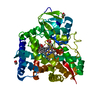 8euhC 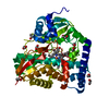 8eulC 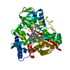 1cptS S: Starting model for refinement C: citing same article ( |
|---|---|
| Similar structure data |
-Links
 PDBj
PDBj
- Assembly
| Deposited unit | 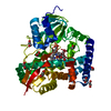
| ||||||||||||
|---|---|---|---|---|---|---|---|---|---|---|---|---|---|
| 1 |
| ||||||||||||
| Unit cell |
|
-Components
| #1: Protein | Mass: 50148.562 Da / Num. of mol.: 1 Source method: isolated from a genetically manipulated source Source: (gene. exp.) Pseudomonas sp. (bacteria) / Gene: cyp108, terPC / Production host: References: UniProt: P33006, Oxidoreductases; Acting on paired donors, with incorporation or reduction of molecular oxygen | ||||
|---|---|---|---|---|---|
| #2: Chemical | ChemComp-HEM /   Mass: 616.487 Da / Num. of mol.: 1 / Source method: obtained synthetically / Formula: C34H32FeN4O4 / Feature type: SUBJECT OF INVESTIGATION Mass: 616.487 Da / Num. of mol.: 1 / Source method: obtained synthetically / Formula: C34H32FeN4O4 / Feature type: SUBJECT OF INVESTIGATION | ||||
| #3: Chemical | ChemComp-EDO /   Mass: 62.068 Da / Num. of mol.: 12 / Source method: obtained synthetically / Formula: C2H6O2 Mass: 62.068 Da / Num. of mol.: 12 / Source method: obtained synthetically / Formula: C2H6O2#4: Water | ChemComp-HOH / |  Mass: 18.015 Da / Num. of mol.: 281 / Source method: isolated from a natural source / Formula: H2O Mass: 18.015 Da / Num. of mol.: 281 / Source method: isolated from a natural source / Formula: H2OHas ligand of interest | Y | |
-Experimental details
-Experiment
| Experiment | Method: X-RAY DIFFRACTION / Number of used crystals: 1 |
|---|
- Sample preparation
| Crystal | Density Matthews: 3.11 Å3/Da / Density % sol: 60.41 % |
|---|---|
| Crystal grow | Temperature: 277 K / Method: vapor diffusion, sitting drop Details: 0.06 M magnesium chloride hexahyrate, 0.06 M calcium chloride dihydrate, 0.1 M sodium HEPES and MOPS (acid) pH 7.5, 20% v/v ethylene glycol, 10% v/v PEG 8000 |
-Data collection
| Diffraction | Mean temperature: 77 K / Serial crystal experiment: N |
|---|---|
| Diffraction source | Source: SYNCHROTRON / Site: ALS / Beamline: 8.2.1 / Wavelength: 1 Å |
| Detector | Type: ADSC QUANTUM 315r / Detector: CCD / Date: Jun 10, 2022 |
| Radiation | Protocol: SINGLE WAVELENGTH / Monochromatic (M) / Laue (L): M / Scattering type: x-ray |
| Radiation wavelength | Wavelength: 1 Å / Relative weight: 1 |
| Reflection | Resolution: 1.98→59.52 Å / Num. obs: 46098 / % possible obs: 99.92 % / Redundancy: 20.1 % / Biso Wilson estimate: 27.62 Å2 / CC1/2: 0.998 / Rpim(I) all: 0.01765 / Net I/σ(I): 27.93 |
| Reflection shell | Resolution: 1.984→2.055 Å / Mean I/σ(I) obs: 11.55 / Num. unique obs: 4465 / CC1/2: 0.995 / Rpim(I) all: 0.04957 / % possible all: 99.98 |
- Processing
| Software |
| ||||||||||||||||||||||||||||||||||||||||||||||||||||||||||||||||||||||||||||||||||||||||||||||||||||||||||||||||||||||||||||||
|---|---|---|---|---|---|---|---|---|---|---|---|---|---|---|---|---|---|---|---|---|---|---|---|---|---|---|---|---|---|---|---|---|---|---|---|---|---|---|---|---|---|---|---|---|---|---|---|---|---|---|---|---|---|---|---|---|---|---|---|---|---|---|---|---|---|---|---|---|---|---|---|---|---|---|---|---|---|---|---|---|---|---|---|---|---|---|---|---|---|---|---|---|---|---|---|---|---|---|---|---|---|---|---|---|---|---|---|---|---|---|---|---|---|---|---|---|---|---|---|---|---|---|---|---|---|---|---|
| Refinement | Method to determine structure: MOLECULAR REPLACEMENT Starting model: 1CPT Resolution: 1.98→59.52 Å / SU ML: 0.154 / Cross valid method: FREE R-VALUE / σ(F): 1.34 / Phase error: 17.8695 Stereochemistry target values: GeoStd + Monomer Library + CDL v1.2
| ||||||||||||||||||||||||||||||||||||||||||||||||||||||||||||||||||||||||||||||||||||||||||||||||||||||||||||||||||||||||||||||
| Solvent computation | Shrinkage radii: 0.9 Å / VDW probe radii: 1.11 Å / Solvent model: FLAT BULK SOLVENT MODEL | ||||||||||||||||||||||||||||||||||||||||||||||||||||||||||||||||||||||||||||||||||||||||||||||||||||||||||||||||||||||||||||||
| Displacement parameters | Biso mean: 35.07 Å2 | ||||||||||||||||||||||||||||||||||||||||||||||||||||||||||||||||||||||||||||||||||||||||||||||||||||||||||||||||||||||||||||||
| Refinement step | Cycle: LAST / Resolution: 1.98→59.52 Å
| ||||||||||||||||||||||||||||||||||||||||||||||||||||||||||||||||||||||||||||||||||||||||||||||||||||||||||||||||||||||||||||||
| Refine LS restraints |
| ||||||||||||||||||||||||||||||||||||||||||||||||||||||||||||||||||||||||||||||||||||||||||||||||||||||||||||||||||||||||||||||
| LS refinement shell |
|
