 Movie
Movie Controller

[English] 日本語
 Yorodumi
- PDB-8esf: Crystal structure of human Nischarin PX and LRR domains with engi... -
+ Open data

- Basic information
| Entry | Database: PDB / ID: 8esf | ||||||
|---|---|---|---|---|---|---|---|
| Title | Crystal structure of human Nischarin PX and LRR domains with engineered mutations | ||||||
 Components Components | Nischarin | ||||||
 Keywords Keywords | ENDOCYTOSIS / Nischarin / PX domain / leucine-rich repeat domain | ||||||
| Function / homology |  Function and homology information Function and homology informationRND2 GTPase cycle / RND3 GTPase cycle / Rac protein signal transduction / intercellular bridge / RAC1 GTPase cycle / phosphatidylinositol binding / negative regulation of cell migration / recycling endosome / integrin binding / microtubule cytoskeleton ...RND2 GTPase cycle / RND3 GTPase cycle / Rac protein signal transduction / intercellular bridge / RAC1 GTPase cycle / phosphatidylinositol binding / negative regulation of cell migration / recycling endosome / integrin binding / microtubule cytoskeleton / actin cytoskeleton organization / early endosome / intracellular membrane-bounded organelle / apoptotic process / nucleoplasm / identical protein binding / membrane / plasma membrane / cytoplasm / cytosol Similarity search - Function | ||||||
| Biological species |  Homo sapiens (human) Homo sapiens (human) | ||||||
| Method |  X-RAY DIFFRACTION / SYNCHROTRON / MOLECULAR REPLACEMENT / Resolution: 2.56 Å X-RAY DIFFRACTION / SYNCHROTRON / MOLECULAR REPLACEMENT / Resolution: 2.56 Å | ||||||
 Authors Authors | Eldershaw, D.E. / Collins, B.M. | ||||||
| Funding support |  Australia, 1items Australia, 1items
| ||||||
 Citation Citation | Journal: To Be Published Title: Crystal structure of human Nischarin PX and LRR domains with engineered mutations Authors: Eldershaw, D.E. / Collins, B.M. | ||||||
| History |
|
- Structure visualization
| Structure viewer | Molecule: MolmilJmol/JSmol |
|---|
- Downloads & links
-Download
| PDBx/mmCIF format | 8esf.cif.gz | 404 KB | Display | PDBx/mmCIF format |
|---|---|---|---|---|
| PDB format | pdb8esf.ent.gz | 268.5 KB | Display | PDB format |
| PDBx/mmJSON format | 8esf.json.gz | Tree view | PDBx/mmJSON format | |
| Others |  Other downloads Other downloads |
-Validation report
| Summary document | 8esf_validation.pdf.gz | 786.7 KB | Display | wwPDB validaton report |
|---|---|---|---|---|
| Full document | 8esf_full_validation.pdf.gz | 793.4 KB | Display | |
| Data in XML | 8esf_validation.xml.gz | 28.7 KB | Display | |
| Data in CIF | 8esf_validation.cif.gz | 39.8 KB | Display | |
| Arichive directory | https://data.pdbj.org/pub/pdb/validation_reports/es/8esfftp://data.pdbj.org/pub/pdb/validation_reports/es/8esf | HTTPS FTP |
-Related structure data
| Similar structure data |
|---|
-Links
 PDBj
PDBj


- Assembly
| Deposited unit | 
| ||||||||||||
|---|---|---|---|---|---|---|---|---|---|---|---|---|---|
| 1 | 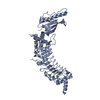
| ||||||||||||
| 2 | 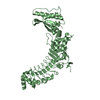
| ||||||||||||
| Unit cell |
|
-Components
| #1: Protein | Mass: 56393.168 Da / Num. of mol.: 2 Source method: isolated from a genetically manipulated source Source: (gene. exp.) Homo sapiens (human) / Gene: NISCH, IRAS, KIAA0975 / Production host:  #2: Chemical | ChemComp-IMD / |   Mass: 69.085 Da / Num. of mol.: 1 / Source method: obtained synthetically / Formula: C3H5N2 / Feature type: SUBJECT OF INVESTIGATION Mass: 69.085 Da / Num. of mol.: 1 / Source method: obtained synthetically / Formula: C3H5N2 / Feature type: SUBJECT OF INVESTIGATION#3: Water | ChemComp-HOH / |  Mass: 18.015 Da / Num. of mol.: 28 / Source method: isolated from a natural source / Formula: H2O Mass: 18.015 Da / Num. of mol.: 28 / Source method: isolated from a natural source / Formula: H2OHas ligand of interest | Y | |
|---|
-Experimental details
-Experiment
| Experiment | Method: X-RAY DIFFRACTION / Number of used crystals: 1 |
|---|
- Sample preparation
| Crystal | Density Matthews: 2.95 Å3/Da / Density % sol: 58.29 % |
|---|---|
| Crystal grow | Temperature: 293 K / Method: vapor diffusion, hanging drop / pH: 8 Details: 0.1 M imidazole, 10% PEG-8000, 0.1 M sodium fluoride |
-Data collection
| Diffraction | Mean temperature: 100 K / Serial crystal experiment: N |
|---|---|
| Diffraction source | Source: SYNCHROTRON / Site: Australian Synchrotron / Beamline: MX2 / Wavelength: 0.9537 Å |
| Detector | Type: DECTRIS EIGER2 X 16M / Detector: PIXEL / Date: Oct 13, 2022 |
| Radiation | Protocol: SINGLE WAVELENGTH / Monochromatic (M) / Laue (L): M / Scattering type: x-ray |
| Radiation wavelength | Wavelength: 0.9537 Å / Relative weight: 1 |
| Reflection | Resolution: 2.56→49.12 Å / Num. obs: 42220 / % possible obs: 99.1 % / Redundancy: 6.9 % / Biso Wilson estimate: 69.77 Å2 / CC1/2: 0.999 / Rpim(I) all: 0.032 / Net I/σ(I): 11.3 |
| Reflection shell | Resolution: 2.56→2.65 Å / Num. unique obs: 4092 / CC1/2: 0.672 |
- Processing
| Software |
| |||||||||||||||||||||||||||||||||||||||||||||||||||||||||||||||||||||||||||||||||||||||||||||||||||||||||
|---|---|---|---|---|---|---|---|---|---|---|---|---|---|---|---|---|---|---|---|---|---|---|---|---|---|---|---|---|---|---|---|---|---|---|---|---|---|---|---|---|---|---|---|---|---|---|---|---|---|---|---|---|---|---|---|---|---|---|---|---|---|---|---|---|---|---|---|---|---|---|---|---|---|---|---|---|---|---|---|---|---|---|---|---|---|---|---|---|---|---|---|---|---|---|---|---|---|---|---|---|---|---|---|---|---|---|
| Refinement | Method to determine structure: MOLECULAR REPLACEMENT Starting model: AlphaFold2 model Resolution: 2.56→43.96 Å / SU ML: 0.3487 / Cross valid method: FREE R-VALUE / σ(F): 1.36 / Phase error: 27.2783 Stereochemistry target values: GeoStd + Monomer Library + CDL v1.2
| |||||||||||||||||||||||||||||||||||||||||||||||||||||||||||||||||||||||||||||||||||||||||||||||||||||||||
| Solvent computation | Shrinkage radii: 0.9 Å / VDW probe radii: 1.11 Å / Solvent model: FLAT BULK SOLVENT MODEL | |||||||||||||||||||||||||||||||||||||||||||||||||||||||||||||||||||||||||||||||||||||||||||||||||||||||||
| Displacement parameters | Biso mean: 76.8 Å2 | |||||||||||||||||||||||||||||||||||||||||||||||||||||||||||||||||||||||||||||||||||||||||||||||||||||||||
| Refinement step | Cycle: LAST / Resolution: 2.56→43.96 Å
| |||||||||||||||||||||||||||||||||||||||||||||||||||||||||||||||||||||||||||||||||||||||||||||||||||||||||
| Refine LS restraints |
| |||||||||||||||||||||||||||||||||||||||||||||||||||||||||||||||||||||||||||||||||||||||||||||||||||||||||
| LS refinement shell |
|
