 Movie
Movie Controller


+ Open data

- Basic information
| Entry | Database: PDB / ID: 8esd | ||||||
|---|---|---|---|---|---|---|---|
| Title | Crystal structure of COMMD7-COMMD9-COMMD5-COMMD10 tetramer | ||||||
 Components Components |
| ||||||
 Keywords Keywords | ENDOCYTOSIS / Complex / Commander / Retriever / Commd5 / Commd7 / Commd9 / Commd10 / Commd | ||||||
| Function / homology |  Function and homology information Function and homology information: / sodium ion transport / NF-kappaB binding / cholesterol homeostasis / tumor necrosis factor-mediated signaling pathway / Neddylation / cytoplasmic vesicle / secretory granule lumen / ficolin-1-rich granule lumen / negative regulation of DNA-templated transcription ...: / sodium ion transport / NF-kappaB binding / cholesterol homeostasis / tumor necrosis factor-mediated signaling pathway / Neddylation / cytoplasmic vesicle / secretory granule lumen / ficolin-1-rich granule lumen / negative regulation of DNA-templated transcription / Neutrophil degranulation / Golgi apparatus / extracellular region / nucleoplasm / nucleus / cytoplasm / cytosol Similarity search - Function | ||||||
| Biological species |  Homo sapiens (human) Homo sapiens (human) | ||||||
| Method |  X-RAY DIFFRACTION / SYNCHROTRON / MOLECULAR REPLACEMENT / Resolution: 3.33 Å X-RAY DIFFRACTION / SYNCHROTRON / MOLECULAR REPLACEMENT / Resolution: 3.33 Å | ||||||
 Authors Authors | Healy, M.D. / Collins, B.M. | ||||||
| Funding support | 1items
| ||||||
 Citation Citation | Journal: Cell / Year: 2023 Title: Structure of the endosomal Commander complex linked to Ritscher-Schinzel syndrome. Authors: Michael D Healy / Kerrie E McNally / Rebeka Butkovič / Molly Chilton / Kohji Kato / Joanna Sacharz / Calum McConville / Edmund R R Moody / Shrestha Shaw / Vicente J Planelles-Herrero / ...Authors: Michael D Healy / Kerrie E McNally / Rebeka Butkovič / Molly Chilton / Kohji Kato / Joanna Sacharz / Calum McConville / Edmund R R Moody / Shrestha Shaw / Vicente J Planelles-Herrero / Sathish K N Yadav / Jennifer Ross / Ufuk Borucu / Catherine S Palmer / Kai-En Chen / Tristan I Croll / Ryan J Hall / Nikeisha J Caruana / Rajesh Ghai / Thi H D Nguyen / Kate J Heesom / Shinji Saitoh / Imre Berger / Christiane Schaffitzel / Tom A Williams / David A Stroud / Emmanuel Derivery / Brett M Collins / Peter J Cullen /    Abstract: The Commander complex is required for endosomal recycling of diverse transmembrane cargos and is mutated in Ritscher-Schinzel syndrome. It comprises two sub-assemblies: Retriever composed of VPS35L, ...The Commander complex is required for endosomal recycling of diverse transmembrane cargos and is mutated in Ritscher-Schinzel syndrome. It comprises two sub-assemblies: Retriever composed of VPS35L, VPS26C, and VPS29; and the CCC complex which contains twelve subunits: COMMD1-COMMD10 and the coiled-coil domain-containing (CCDC) proteins CCDC22 and CCDC93. Combining X-ray crystallography, electron cryomicroscopy, and in silico predictions, we have assembled a complete structural model of Commander. Retriever is distantly related to the endosomal Retromer complex but has unique features preventing the shared VPS29 subunit from interacting with Retromer-associated factors. The COMMD proteins form a distinctive hetero-decameric ring stabilized by extensive interactions with CCDC22 and CCDC93. These adopt a coiled-coil structure that connects the CCC and Retriever assemblies and recruits a 16th subunit, DENND10, to form the complete Commander complex. The structure allows mapping of disease-causing mutations and reveals the molecular features required for the function of this evolutionarily conserved trafficking machinery. | ||||||
| History |
|
- Structure visualization
| Structure viewer | Molecule: MolmilJmol/JSmol |
|---|
- Downloads & links
-Download
| PDBx/mmCIF format | 8esd.cif.gz | 118.5 KB | Display | PDBx/mmCIF format |
|---|---|---|---|---|
| PDB format | pdb8esd.ent.gz | 89.5 KB | Display | PDB format |
| PDBx/mmJSON format | 8esd.json.gz | Tree view | PDBx/mmJSON format | |
| Others |  Other downloads Other downloads |
-Validation report
| Arichive directory | https://data.pdbj.org/pub/pdb/validation_reports/es/8esdftp://data.pdbj.org/pub/pdb/validation_reports/es/8esd | HTTPS FTP |
|---|
-Related structure data
| Related structure data |  8eseC 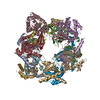 8f2rC 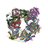 8f2uC  6bp6S S: Starting model for refinement C: citing same article ( |
|---|---|
| Similar structure data |


-Links
 PDBj
PDBj- Assembly
| Deposited unit | 
| ||||||||||||
|---|---|---|---|---|---|---|---|---|---|---|---|---|---|
| 1 |
| ||||||||||||
| Unit cell |
|
-Components
| #1: Protein | Mass: 21828.939 Da / Num. of mol.: 1 Source method: isolated from a genetically manipulated source Source: (gene. exp.) Homo sapiens (human) / Gene: COMMD10, HSPC305, PTD002 / Production host:  |
|---|---|
| #2: Protein | Mass: 21457.607 Da / Num. of mol.: 1 Source method: isolated from a genetically manipulated source Source: (gene. exp.) Homo sapiens (human) / Gene: COMMD9, HSPC166 / Production host: |
| #3: Protein | Mass: 8636.016 Da / Num. of mol.: 1 Source method: isolated from a genetically manipulated source Source: (gene. exp.) Homo sapiens (human) / Gene: COMMD5, HT002 / Production host: |
| #4: Protein | Mass: 8172.452 Da / Num. of mol.: 1 Source method: isolated from a genetically manipulated source Source: (gene. exp.) Homo sapiens (human) / Gene: COMMD7, C20orf92 / Production host: |
-Experimental details
-Experiment
| Experiment | Method: X-RAY DIFFRACTION / Number of used crystals: 1 |
|---|
- Sample preparation
| Crystal | Density Matthews: 4.37 Å3/Da / Density % sol: 71.82 % |
|---|---|
| Crystal grow | Temperature: 293.15 K / Method: vapor diffusion, hanging drop Details: 2 uM crown ether and 10% glycerol and grown in 22% ethanol and 5 mM EDTA |
-Data collection
| Diffraction | Mean temperature: 100 K / Serial crystal experiment: N |
|---|---|
| Diffraction source | Source: SYNCHROTRON / Site: Australian Synchrotron / Beamline: MX2 / Wavelength: 0.95372 Å |
| Detector | Type: DECTRIS EIGER X 16M / Detector: PIXEL / Date: Oct 13, 2022 |
| Radiation | Protocol: SINGLE WAVELENGTH / Monochromatic (M) / Laue (L): M / Scattering type: x-ray |
| Radiation wavelength | Wavelength: 0.95372 Å / Relative weight: 1 |
| Reflection | Resolution: 3.13→48.52 Å / Num. obs: 16930 / % possible obs: 96.5 % / Redundancy: 5.1 % / Biso Wilson estimate: 103.34 Å2 / CC1/2: 0.997 / Rpim(I) all: 0.08 / Net I/σ(I): 6.2 |
| Reflection shell | Resolution: 3.13→3.35 Å / Mean I/σ(I) obs: 0.9 / Num. unique obs: 2673 / CC1/2: 0.56 / Rpim(I) all: 0.908 |
- Processing
| Software |
| |||||||||||||||||||||||||||||||||||||||||||||||||||||||||||||||||||||||||||||
|---|---|---|---|---|---|---|---|---|---|---|---|---|---|---|---|---|---|---|---|---|---|---|---|---|---|---|---|---|---|---|---|---|---|---|---|---|---|---|---|---|---|---|---|---|---|---|---|---|---|---|---|---|---|---|---|---|---|---|---|---|---|---|---|---|---|---|---|---|---|---|---|---|---|---|---|---|---|---|
| Refinement | Method to determine structure: MOLECULAR REPLACEMENT Starting model: 6BP6 Resolution: 3.33→38.5 Å / SU ML: 0.4188 / Cross valid method: FREE R-VALUE / σ(F): 0 / Phase error: 31.9287 Stereochemistry target values: GeoStd + Monomer Library + CDL v1.2
| |||||||||||||||||||||||||||||||||||||||||||||||||||||||||||||||||||||||||||||
| Solvent computation | Shrinkage radii: 0.9 Å / VDW probe radii: 1.11 Å / Solvent model: FLAT BULK SOLVENT MODEL | |||||||||||||||||||||||||||||||||||||||||||||||||||||||||||||||||||||||||||||
| Displacement parameters | Biso mean: 127.07 Å2 | |||||||||||||||||||||||||||||||||||||||||||||||||||||||||||||||||||||||||||||
| Refinement step | Cycle: LAST / Resolution: 3.33→38.5 Å
| |||||||||||||||||||||||||||||||||||||||||||||||||||||||||||||||||||||||||||||
| Refine LS restraints |
| |||||||||||||||||||||||||||||||||||||||||||||||||||||||||||||||||||||||||||||
| LS refinement shell |
|
