 Movie
Movie Controller

[English] 日本語
 Yorodumi
- PDB-8eqm: Structure of a dimeric photosystem II complex acclimated to far-r... -
+ Open data

- Basic information
| Entry | Database: PDB / ID: 8eqm | ||||||||||||
|---|---|---|---|---|---|---|---|---|---|---|---|---|---|
| Title | Structure of a dimeric photosystem II complex acclimated to far-red light | ||||||||||||
 Components Components |
| ||||||||||||
 Keywords Keywords | PHOTOSYNTHESIS / Far-red / photosystem II / cyanobacteria / chlorophyll | ||||||||||||
| Function / homology |  Function and homology information Function and homology information: / photosystem II oxygen evolving complex / photosystem II assembly / oxygen evolving activity / photosystem II stabilization / photosystem II reaction center / photosystem II / photosynthetic electron transport chain / photosystem II / extrinsic component of membrane ...: / photosystem II oxygen evolving complex / photosystem II assembly / oxygen evolving activity / photosystem II stabilization / photosystem II reaction center / photosystem II / photosynthetic electron transport chain / photosystem II / extrinsic component of membrane / plasma membrane-derived thylakoid membrane / photosynthetic electron transport in photosystem II / chlorophyll binding / photosynthesis, light reaction / phosphate ion binding / photosynthesis / respiratory electron transport chain / electron transfer activity / protein stabilization / iron ion binding / heme binding / metal ion binding Similarity search - Function | ||||||||||||
| Biological species |  Synechococcus sp. PCC 7335 (bacteria) Synechococcus sp. PCC 7335 (bacteria) | ||||||||||||
| Method | ELECTRON MICROSCOPY / single particle reconstruction / cryo EM / Resolution: 2.6 Å | ||||||||||||
 Authors Authors | Gisriel, C.J. / Shen, G. / Flesher, D.A. / Kurashov, V. / Golbeck, J.H. / Brudvig, G.W. / Amin, M. / Bryant, D.A. | ||||||||||||
| Funding support |  United States, 3items United States, 3items
| ||||||||||||
 Citation Citation | Journal: J Biol Chem / Year: 2023 Title: Structure of a dimeric photosystem II complex from a cyanobacterium acclimated to far-red light. Authors: Christopher J Gisriel / Gaozhong Shen / David A Flesher / Vasily Kurashov / John H Golbeck / Gary W Brudvig / Muhamed Amin / Donald A Bryant /  Abstract: Photosystem II (PSII) is the water-splitting enzyme central to oxygenic photosynthesis. To drive water oxidation, light is harvested by accessory pigments, mostly chlorophyll (Chl) a molecules, which ...Photosystem II (PSII) is the water-splitting enzyme central to oxygenic photosynthesis. To drive water oxidation, light is harvested by accessory pigments, mostly chlorophyll (Chl) a molecules, which absorb visible light (400-700 nm). Some cyanobacteria facultatively acclimate to shaded environments by altering their photosynthetic machinery to additionally absorb far-red light (FRL, 700-800 nm), a process termed far-red light photoacclimation or FaRLiP. During far-red light photoacclimation, FRL-PSII is assembled with FRL-specific isoforms of the subunits PsbA, PsbB, PsbC, PsbD, and PsbH, and some Chl-binding sites contain Chls d or f instead of the usual Chl a. The structure of an apo-FRL-PSII monomer lacking the FRL-specific PsbH subunit has previously been determined, but visualization of the dimeric complex has remained elusive. Here, we report the cryo-EM structure of a dimeric FRL-PSII complex. The site assignments for Chls d and f are consistent with those assigned in the previous apo-FRL-PSII monomeric structure. All sites that bind Chl d or Chl f at high occupancy exhibit a FRL-specific interaction of the formyl moiety of the Chl d or Chl f with the protein environment, which in some cases involves a phenylalanine sidechain. The structure retains the FRL-specific PsbH2 subunit, which appears to alter the energetic landscape of FRL-PSII, redirecting energy transfer from the phycobiliprotein complex to a Chl f molecule bound by PsbB2 that acts as a bridge for energy transfer to the electron transfer chain. Collectively, these observations extend our previous understanding of the structure-function relationship that allows PSII to function using lower energy FRL. | ||||||||||||
| History |
|
- Structure visualization
| Structure viewer | Molecule: MolmilJmol/JSmol |
|---|
- Downloads & links
-Download
| PDBx/mmCIF format | 8eqm.cif.gz | 970.8 KB | Display | PDBx/mmCIF format |
|---|---|---|---|---|
| PDB format | pdb8eqm.ent.gz | 805.3 KB | Display | PDB format |
| PDBx/mmJSON format | 8eqm.json.gz | Tree view | PDBx/mmJSON format | |
| Others |  Other downloads Other downloads |
-Validation report
| Arichive directory | https://data.pdbj.org/pub/pdb/validation_reports/eq/8eqmftp://data.pdbj.org/pub/pdb/validation_reports/eq/8eqm | HTTPS FTP |
|---|
-Related structure data
| Related structure data | 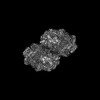 28539MC M: map data used to model this data C: citing same article ( |
|---|---|
| Similar structure data |
-Links
 PDBj
PDBj


- Assembly
| Deposited unit | 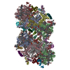
|
|---|---|
| 1 |
|
-Components
-Photosystem II ... , 13 types, 26 molecules AaBbCcDdHhIiKkLlMmOoTtUuXx
| #1: Protein | Mass: 39958.668 Da / Num. of mol.: 2 / Source method: isolated from a natural source / Source: (natural) Synechococcus sp. PCC 7335 (bacteria) / Strain: ATCC 29403 / PCC 7335#2: Protein | Mass: 56440.980 Da / Num. of mol.: 2 / Source method: isolated from a natural source / Source: (natural) Synechococcus sp. PCC 7335 (bacteria) / Strain: ATCC 29403 / PCC 7335 / References: UniProt: B4WKI1#3: Protein | Mass: 52375.664 Da / Num. of mol.: 2 / Source method: isolated from a natural source / Source: (natural) Synechococcus sp. PCC 7335 (bacteria) / Strain: ATCC 29403 / PCC 7335 / References: UniProt: B4WKI2#4: Protein | Mass: 39596.496 Da / Num. of mol.: 2 / Source method: isolated from a natural source / Source: (natural) Synechococcus sp. PCC 7335 (bacteria) / Strain: ATCC 29403 / PCC 7335 / References: UniProt: B4WKI3#7: Protein | Mass: 7247.715 Da / Num. of mol.: 2 / Source method: isolated from a natural source / Source: (natural) Synechococcus sp. PCC 7335 (bacteria) / Strain: ATCC 29403 / PCC 7335 / References: UniProt: B4WKI0#8: Protein/peptide | Mass: 4229.917 Da / Num. of mol.: 2 / Source method: isolated from a natural source / Source: (natural) Synechococcus sp. PCC 7335 (bacteria) / Strain: ATCC 29403 / PCC 7335 / References: UniProt: B4WM03#9: Protein/peptide | Mass: 5069.053 Da / Num. of mol.: 2 / Source method: isolated from a natural source / Source: (natural) Synechococcus sp. PCC 7335 (bacteria) / Strain: ATCC 29403 / PCC 7335 / References: UniProt: B4WR12#10: Protein/peptide | Mass: 4566.261 Da / Num. of mol.: 2 / Source method: isolated from a natural source / Source: (natural) Synechococcus sp. PCC 7335 (bacteria) / Strain: ATCC 29403 / PCC 7335 / References: UniProt: B4WII3#11: Protein/peptide | Mass: 3980.772 Da / Num. of mol.: 2 / Source method: isolated from a natural source / Source: (natural) Synechococcus sp. PCC 7335 (bacteria) / Strain: ATCC 29403 / PCC 7335#12: Protein | Mass: 29319.096 Da / Num. of mol.: 2 / Source method: isolated from a natural source / Source: (natural) Synechococcus sp. PCC 7335 (bacteria) / Strain: ATCC 29403 / PCC 7335 / References: UniProt: B4WKJ1#13: Protein/peptide | Mass: 3606.362 Da / Num. of mol.: 2 / Source method: isolated from a natural source / Source: (natural) Synechococcus sp. PCC 7335 (bacteria) / Strain: ATCC 29403 / PCC 7335 / References: UniProt: A0A2W4UG77#14: Protein | Mass: 17325.834 Da / Num. of mol.: 2 / Source method: isolated from a natural source / Source: (natural) Synechococcus sp. PCC 7335 (bacteria) / Strain: ATCC 29403 / PCC 7335 / References: UniProt: B4WMT5#16: Protein/peptide | Mass: 4273.182 Da / Num. of mol.: 2 / Source method: isolated from a natural source / Source: (natural) Synechococcus sp. PCC 7335 (bacteria) / Strain: ATCC 29403 / PCC 7335 / References: UniProt: B4WIZ7 |
|---|
-Cytochrome b559 subunit ... , 2 types, 4 molecules EeFf
| #5: Protein | Mass: 9136.276 Da / Num. of mol.: 2 / Source method: isolated from a natural source / Source: (natural) Synechococcus sp. PCC 7335 (bacteria) / Strain: ATCC 29403 / PCC 7335 / References: UniProt: B4WII1#6: Protein/peptide | Mass: 5023.960 Da / Num. of mol.: 2 / Source method: isolated from a natural source / Source: (natural) Synechococcus sp. PCC 7335 (bacteria) / Strain: ATCC 29403 / PCC 7335 / References: UniProt: B4WKJ2 |
|---|
-Protein , 1 types, 2 molecules Vv
| #15: Protein | Mass: 18814.387 Da / Num. of mol.: 2 / Source method: isolated from a natural source / Source: (natural) Synechococcus sp. PCC 7335 (bacteria) / Strain: ATCC 29403 / PCC 7335 / References: UniProt: B4WI32 |
|---|
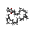
-Sugars , 2 types, 8 molecules 

| #28: Sugar | ChemComp-DGD /  Type: saccharide / Mass: 949.299 Da / Num. of mol.: 6 / Source method: obtained synthetically / Formula: C51H96O15 Type: saccharide / Mass: 949.299 Da / Num. of mol.: 6 / Source method: obtained synthetically / Formula: C51H96O15#33: Sugar |  Type: D-saccharide / Mass: 510.615 Da / Num. of mol.: 2 / Source method: obtained synthetically / Formula: C24H46O11 / Comment: detergent*YM Type: D-saccharide / Mass: 510.615 Da / Num. of mol.: 2 / Source method: obtained synthetically / Formula: C24H46O11 / Comment: detergent*YM |
|---|
-Non-polymers , 16 types, 714 molecules 


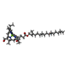


| #17: Chemical |  Mass: 339.827 Da / Num. of mol.: 2 / Source method: obtained synthetically / Formula: CaMn4O5 Mass: 339.827 Da / Num. of mol.: 2 / Source method: obtained synthetically / Formula: CaMn4O5#18: Chemical |  Mass: 55.845 Da / Num. of mol.: 2 / Source method: obtained synthetically / Formula: Fe Mass: 55.845 Da / Num. of mol.: 2 / Source method: obtained synthetically / Formula: Fe#19: Chemical | ChemComp-CL /  Mass: 35.453 Da / Num. of mol.: 4 / Source method: obtained synthetically / Formula: Cl Mass: 35.453 Da / Num. of mol.: 4 / Source method: obtained synthetically / Formula: Cl#20: Chemical | ChemComp-CLA /  Mass: 893.489 Da / Num. of mol.: 60 / Source method: obtained synthetically / Formula: C55H72MgN4O5 Mass: 893.489 Da / Num. of mol.: 60 / Source method: obtained synthetically / Formula: C55H72MgN4O5#21: Chemical | ChemComp-PHO /  Mass: 871.200 Da / Num. of mol.: 4 / Source method: obtained synthetically / Formula: C55H74N4O5 Mass: 871.200 Da / Num. of mol.: 4 / Source method: obtained synthetically / Formula: C55H74N4O5#22: Chemical | ChemComp-BCR /  Mass: 536.873 Da / Num. of mol.: 14 / Source method: obtained synthetically / Formula: C40H56 Mass: 536.873 Da / Num. of mol.: 14 / Source method: obtained synthetically / Formula: C40H56#23: Chemical | ChemComp-SQD /  Mass: 795.116 Da / Num. of mol.: 4 / Source method: obtained synthetically / Formula: C41H78O12S Mass: 795.116 Da / Num. of mol.: 4 / Source method: obtained synthetically / Formula: C41H78O12S#24: Chemical | ChemComp-PL9 /  Mass: 749.201 Da / Num. of mol.: 4 / Source method: obtained synthetically / Formula: C53H80O2 Mass: 749.201 Da / Num. of mol.: 4 / Source method: obtained synthetically / Formula: C53H80O2#25: Chemical |  Mass: 61.017 Da / Num. of mol.: 2 / Source method: obtained synthetically / Formula: CHO3 / Comment: pH buffer*YM Mass: 61.017 Da / Num. of mol.: 2 / Source method: obtained synthetically / Formula: CHO3 / Comment: pH buffer*YM#26: Chemical | ChemComp-LHG /  Mass: 722.970 Da / Num. of mol.: 8 / Source method: obtained synthetically / Formula: C38H75O10P / Comment: phospholipid*YM Mass: 722.970 Da / Num. of mol.: 8 / Source method: obtained synthetically / Formula: C38H75O10P / Comment: phospholipid*YM#27: Chemical | ChemComp-F6C /  Mass: 905.457 Da / Num. of mol.: 8 / Source method: obtained synthetically / Formula: C55H68MgN4O6 / Feature type: SUBJECT OF INVESTIGATION Mass: 905.457 Da / Num. of mol.: 8 / Source method: obtained synthetically / Formula: C55H68MgN4O6 / Feature type: SUBJECT OF INVESTIGATION#29: Chemical |  Mass: 895.462 Da / Num. of mol.: 2 / Source method: obtained synthetically / Formula: C54H70MgN4O6 / Feature type: SUBJECT OF INVESTIGATION Mass: 895.462 Da / Num. of mol.: 2 / Source method: obtained synthetically / Formula: C54H70MgN4O6 / Feature type: SUBJECT OF INVESTIGATION#30: Chemical | ChemComp-LMG /  Mass: 787.158 Da / Num. of mol.: 4 / Source method: obtained synthetically / Formula: C45H86O10 Mass: 787.158 Da / Num. of mol.: 4 / Source method: obtained synthetically / Formula: C45H86O10#31: Chemical | ChemComp-HEM /  Mass: 616.487 Da / Num. of mol.: 4 / Source method: obtained synthetically / Formula: C34H32FeN4O4 Mass: 616.487 Da / Num. of mol.: 4 / Source method: obtained synthetically / Formula: C34H32FeN4O4#32: Chemical |  Mass: 552.872 Da / Num. of mol.: 2 / Source method: obtained synthetically / Formula: C40H56O Mass: 552.872 Da / Num. of mol.: 2 / Source method: obtained synthetically / Formula: C40H56O#34: Water | ChemComp-HOH / | Mass: 18.015 Da / Num. of mol.: 590 / Source method: isolated from a natural source / Formula: H2O |
|---|
-Details
| Has ligand of interest | Y |
|---|---|
| Has protein modification | N |
-Experimental details
-Experiment
| Experiment | Method: ELECTRON MICROSCOPY |
|---|---|
| EM experiment | Aggregation state: PARTICLE / 3D reconstruction method: single particle reconstruction |
- Sample preparation
| Component | Name: Far-red light-acclimated photosystem II / Type: COMPLEX / Entity ID: #3, #1-#2, #4-#16 / Source: NATURAL |
|---|---|
| Source (natural) | Organism: Synechococcus sp. PCC 7335 (bacteria) |
| Buffer solution | pH: 6.5 |
| Specimen | Conc.: 0.65 mg/ml / Embedding applied: NO / Shadowing applied: NO / Staining applied: NO / Vitrification applied: YES |
| Vitrification | Cryogen name: ETHANE |
- Electron microscopy imaging
| Experimental equipment |  Model: Titan Krios / Image courtesy: FEI Company |
|---|---|
| Microscopy | Model: FEI TITAN KRIOS |
| Electron gun | Electron source:  FIELD EMISSION GUN / Accelerating voltage: 300 kV / Illumination mode: FLOOD BEAM FIELD EMISSION GUN / Accelerating voltage: 300 kV / Illumination mode: FLOOD BEAM |
| Electron lens | Mode: BRIGHT FIELD / Nominal defocus max: 2000 nm / Nominal defocus min: 1200 nm |
| Image recording | Electron dose: 41.1 e/Å2 / Film or detector model: GATAN K3 (6k x 4k) |
- Processing
| CTF correction | Type: PHASE FLIPPING AND AMPLITUDE CORRECTION |
|---|---|
| 3D reconstruction | Resolution: 2.6 Å / Resolution method: FSC 0.143 CUT-OFF / Num. of particles: 90191 / Symmetry type: POINT |
