 Movie
Movie Controller

[English] 日本語
 Yorodumi
- PDB-8epz: Crystal structure of Fe-S cluster-dependent dehydratase from Para... -
+ Open data

- Basic information
| Entry | Database: PDB / ID: 8epz | ||||||
|---|---|---|---|---|---|---|---|
| Title | Crystal structure of Fe-S cluster-dependent dehydratase from Paralcaligenes ureilyticus in complex with Mn | ||||||
 Components Components | Dihydroxyacid dehydratase | ||||||
 Keywords Keywords | LYASE / Fe-S dehydratase / sugar-acid dehydratase / ilvD/EDD family / DHAD | ||||||
| Function / homology |  Function and homology information Function and homology informationhydro-lyase activity / 2 iron, 2 sulfur cluster binding / metal ion binding Similarity search - Function | ||||||
| Biological species |  Paralcaligenes ureilyticus (bacteria) Paralcaligenes ureilyticus (bacteria) | ||||||
| Method |  X-RAY DIFFRACTION / SYNCHROTRON / MOLECULAR REPLACEMENT / Resolution: 2.6 Å X-RAY DIFFRACTION / SYNCHROTRON / MOLECULAR REPLACEMENT / Resolution: 2.6 Å | ||||||
 Authors Authors | Bayaraa, T. / Lonhienne, T. / Guddat, L.W. | ||||||
| Funding support |  Australia, 1items Australia, 1items
| ||||||
 Citation Citation | Journal: To Be Published Title: Crystal structure of Fe-S cluster-dependent dehydratase from Paralcaligenes ureilyticus in complex with Mg Authors: Bayaraa, T. / Lonhienne, T. / Guddat, L.W. | ||||||
| History |
|
- Structure visualization
| Structure viewer | Molecule: MolmilJmol/JSmol |
|---|
- Downloads & links
-Download
| PDBx/mmCIF format | 8epz.cif.gz | 224.4 KB | Display | PDBx/mmCIF format |
|---|---|---|---|---|
| PDB format | pdb8epz.ent.gz | 177.8 KB | Display | PDB format |
| PDBx/mmJSON format | 8epz.json.gz | Tree view | PDBx/mmJSON format | |
| Others |  Other downloads Other downloads |
-Validation report
| Summary document | 8epz_validation.pdf.gz | 2.4 MB | Display | wwPDB validaton report |
|---|---|---|---|---|
| Full document | 8epz_full_validation.pdf.gz | 2.5 MB | Display | |
| Data in XML | 8epz_validation.xml.gz | 43 KB | Display | |
| Data in CIF | 8epz_validation.cif.gz | 58 KB | Display | |
| Arichive directory | https://data.pdbj.org/pub/pdb/validation_reports/ep/8epzftp://data.pdbj.org/pub/pdb/validation_reports/ep/8epz | HTTPS FTP |
-Related structure data
| Related structure data |  8ej0S S: Starting model for refinement |
|---|---|
| Similar structure data |
-Links
 PDBj
PDBj


- Assembly
| Deposited unit | 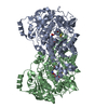
| ||||||||
|---|---|---|---|---|---|---|---|---|---|
| 1 |
| ||||||||
| Unit cell |
|
-Components
-Protein , 1 types, 2 molecules AB
| #1: Protein | Mass: 62651.746 Da / Num. of mol.: 2 Source method: isolated from a genetically manipulated source Source: (gene. exp.) Paralcaligenes ureilyticus (bacteria) / Gene: EDC26_11947 / Production host: |
|---|
-Non-polymers , 5 types, 78 molecules 


| #2: Chemical |  Mass: 175.820 Da / Num. of mol.: 2 / Source method: obtained synthetically / Formula: Fe2S2 / Feature type: SUBJECT OF INVESTIGATION Mass: 175.820 Da / Num. of mol.: 2 / Source method: obtained synthetically / Formula: Fe2S2 / Feature type: SUBJECT OF INVESTIGATION#3: Chemical |  Mass: 54.938 Da / Num. of mol.: 2 / Source method: obtained synthetically / Formula: Mn / Feature type: SUBJECT OF INVESTIGATION Mass: 54.938 Da / Num. of mol.: 2 / Source method: obtained synthetically / Formula: Mn / Feature type: SUBJECT OF INVESTIGATION#4: Chemical |  Mass: 44.010 Da / Num. of mol.: 3 / Source method: obtained synthetically / Formula: CO2 / Feature type: SUBJECT OF INVESTIGATION Mass: 44.010 Da / Num. of mol.: 3 / Source method: obtained synthetically / Formula: CO2 / Feature type: SUBJECT OF INVESTIGATION#5: Chemical | ChemComp-BCT / |  Mass: 61.017 Da / Num. of mol.: 1 / Source method: obtained synthetically / Formula: CHO3 / Feature type: SUBJECT OF INVESTIGATION Mass: 61.017 Da / Num. of mol.: 1 / Source method: obtained synthetically / Formula: CHO3 / Feature type: SUBJECT OF INVESTIGATION#6: Water | ChemComp-HOH / | Mass: 18.015 Da / Num. of mol.: 70 / Source method: isolated from a natural source / Formula: H2O |
|---|
-Details
| Has ligand of interest | Y |
|---|
-Experimental details
-Experiment
| Experiment | Method: X-RAY DIFFRACTION / Number of used crystals: 1 |
|---|
- Sample preparation
| Crystal | Density Matthews: 1.99 Å3/Da / Density % sol: 38.34 % |
|---|---|
| Crystal grow | Temperature: 293 K / Method: vapor diffusion, hanging drop / Details: 0.2 M succinic acid, 15% PEG 3350 |
-Data collection
| Diffraction | Mean temperature: 100 K / Serial crystal experiment: N |
|---|---|
| Diffraction source | Source: SYNCHROTRON / Site: Australian Synchrotron / Beamline: MX1 / Wavelength: 0.954 Å |
| Detector | Type: DECTRIS PILATUS 2M / Detector: PIXEL / Date: Apr 21, 2021 |
| Radiation | Protocol: SINGLE WAVELENGTH / Monochromatic (M) / Laue (L): M / Scattering type: x-ray |
| Radiation wavelength | Wavelength: 0.954 Å / Relative weight: 1 |
| Reflection | Resolution: 2.6→48.107 Å / Num. obs: 44640 / % possible obs: 98.8 % / Redundancy: 5.3 % / Rmerge(I) obs: 0.108 / Rpim(I) all: 0.046 / Net I/σ(I): 9.1 |
| Reflection shell | Resolution: 2.6→2.987 Å / Rmerge(I) obs: 0.747 / Mean I/σ(I) obs: 1.9 / Num. unique obs: 3904 / Rpim(I) all: 0.316 |
- Processing
| Software |
| ||||||||||||||||||||||||||||||||||||||||||||||||||||||||||||||||||
|---|---|---|---|---|---|---|---|---|---|---|---|---|---|---|---|---|---|---|---|---|---|---|---|---|---|---|---|---|---|---|---|---|---|---|---|---|---|---|---|---|---|---|---|---|---|---|---|---|---|---|---|---|---|---|---|---|---|---|---|---|---|---|---|---|---|---|---|
| Refinement | Method to determine structure: MOLECULAR REPLACEMENT Starting model: 8EJ0 Resolution: 2.6→48.107 Å / SU ML: 0.3 / Cross valid method: THROUGHOUT / σ(F): 1.38 / Phase error: 32.35 / Stereochemistry target values: ML
| ||||||||||||||||||||||||||||||||||||||||||||||||||||||||||||||||||
| Solvent computation | Shrinkage radii: 0.9 Å / VDW probe radii: 1.11 Å / Solvent model: FLAT BULK SOLVENT MODEL | ||||||||||||||||||||||||||||||||||||||||||||||||||||||||||||||||||
| Displacement parameters | Biso max: 107.05 Å2 / Biso mean: 61.2977 Å2 / Biso min: 30 Å2 | ||||||||||||||||||||||||||||||||||||||||||||||||||||||||||||||||||
| Refinement step | Cycle: final / Resolution: 2.6→48.107 Å
| ||||||||||||||||||||||||||||||||||||||||||||||||||||||||||||||||||
| Refine LS restraints |
| ||||||||||||||||||||||||||||||||||||||||||||||||||||||||||||||||||
| LS refinement shell | Refine-ID: X-RAY DIFFRACTION / Rfactor Rfree error: 0
|
