 Movie
Movie Controller

[English] 日本語
 Yorodumi
- PDB-8epu: 1.6 A crystal structure of the lipocalin dog allergen Can f 1 wit... -
+ Open data

- Basic information
| Entry | Database: PDB / ID: 8epu | ||||||
|---|---|---|---|---|---|---|---|
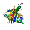
| Title | 1.6 A crystal structure of the lipocalin dog allergen Can f 1 with the C118S mutation | ||||||
 Components Components | Major allergen Can f 1 | ||||||
 Keywords Keywords | ALLERGEN / Lipocalin / Fatty acid binding protein / Calyx | ||||||
| Function / homology | von Ebner's gland protein/ Bos/Can allergen / Lipocalin / small molecule binding / Lipocalin / cytosolic fatty-acid binding protein family / Lipocalin/cytosolic fatty-acid binding domain / Calycin / extracellular space / Major allergen Can f 1 Function and homology information Function and homology information | ||||||
| Biological species |  | ||||||
| Method |  X-RAY DIFFRACTION / SYNCHROTRON / MOLECULAR REPLACEMENT / Resolution: 1.6 Å X-RAY DIFFRACTION / SYNCHROTRON / MOLECULAR REPLACEMENT / Resolution: 1.6 Å | ||||||
 Authors Authors | Min, J. / Pedersen, L.C. / Geoffrey, M.A. | ||||||
| Funding support |  United States, 1items United States, 1items
| ||||||
 Citation Citation | Journal: Front Allergy / Year: 2023 Title: Structural and ligand binding analysis of the pet allergens Can f 1 and Fel d 7. Authors: Min, J. / Foo, A.C.Y. / Gabel, S.A. / Perera, L. / DeRose, E.F. / Pomes, A. / Pedersen, L.C. / Mueller, G.A. | ||||||
| History |
|
- Structure visualization
| Structure viewer | Molecule: MolmilJmol/JSmol |
|---|
- Downloads & links
-Download
| PDBx/mmCIF format | 8epu.cif.gz | 113 KB | Display | PDBx/mmCIF format |
|---|---|---|---|---|
| PDB format | pdb8epu.ent.gz | Display | PDB format | |
| PDBx/mmJSON format | 8epu.json.gz | Tree view | PDBx/mmJSON format | |
| Others |  Other downloads Other downloads |
-Validation report
| Arichive directory | https://data.pdbj.org/pub/pdb/validation_reports/ep/8epuftp://data.pdbj.org/pub/pdb/validation_reports/ep/8epu | HTTPS FTP |
|---|
-Related structure data
-Links
 PDBj
PDBj

- Assembly
| Deposited unit | 
| ||||||||||||
|---|---|---|---|---|---|---|---|---|---|---|---|---|---|
| 1 |
| ||||||||||||
| Unit cell |
| ||||||||||||
| Components on special symmetry positions |
| ||||||||||||
| Details | The oligomeric state is unknown. Lipocalin proteins are often in a transient monomer to oligomer state. |
-Components
| #1: Protein | Mass: 16569.699 Da / Num. of mol.: 1 / Mutation: C118S Source method: isolated from a genetically manipulated source Source: (gene. exp.)  |
|---|---|
| #2: Water | ChemComp-HOH /  Mass: 18.015 Da / Num. of mol.: 68 / Source method: isolated from a natural source / Formula: H2O Mass: 18.015 Da / Num. of mol.: 68 / Source method: isolated from a natural source / Formula: H2O |
| Has protein modification | Y |
-Experimental details
-Experiment
| Experiment | Method: X-RAY DIFFRACTION / Number of used crystals: 1 |
|---|
- Sample preparation
| Crystal | Density Matthews: 1.95 Å3/Da / Density % sol: 37.05 % |
|---|---|
| Crystal grow | Temperature: 293 K / Method: vapor diffusion, hanging drop / Details: 35 % PEG 4000 |
-Data collection
| Diffraction | Mean temperature: 100 K / Serial crystal experiment: N |
|---|---|
| Diffraction source | Source: SYNCHROTRON / Site: APS / Beamline: 22-ID / Wavelength: 1 Å |
| Detector | Type: DECTRIS EIGER X 16M / Detector: PIXEL / Date: Sep 18, 2021 |
| Radiation | Protocol: SINGLE WAVELENGTH / Monochromatic (M) / Laue (L): M / Scattering type: x-ray |
| Radiation wavelength | Wavelength: 1 Å / Relative weight: 1 |
| Reflection | Resolution: 1.6→50 Å / Num. obs: 16064 / % possible obs: 94 % / Redundancy: 3.1 % / Biso Wilson estimate: 24.85 Å2 / CC1/2: 0.986 / Net I/σ(I): 25.98 |
| Reflection shell | Resolution: 1.6→1.63 Å / Num. unique obs: 684 / CC1/2: 0.975 |
- Processing
| Software |
| |||||||||||||||||||||||||||||||||||||||||||||||||||||||||||||||||||||||||||||||||||||||||||||||||||||||||||||||||||||||||||||
|---|---|---|---|---|---|---|---|---|---|---|---|---|---|---|---|---|---|---|---|---|---|---|---|---|---|---|---|---|---|---|---|---|---|---|---|---|---|---|---|---|---|---|---|---|---|---|---|---|---|---|---|---|---|---|---|---|---|---|---|---|---|---|---|---|---|---|---|---|---|---|---|---|---|---|---|---|---|---|---|---|---|---|---|---|---|---|---|---|---|---|---|---|---|---|---|---|---|---|---|---|---|---|---|---|---|---|---|---|---|---|---|---|---|---|---|---|---|---|---|---|---|---|---|---|---|---|
| Refinement | Method to determine structure: MOLECULAR REPLACEMENT Starting model: Alphafold Resolution: 1.6→31.34 Å / SU ML: 0.1854 / Cross valid method: FREE R-VALUE / σ(F): 1.41 / Phase error: 29.3045 Stereochemistry target values: GeoStd + Monomer Library + CDL v1.2
| |||||||||||||||||||||||||||||||||||||||||||||||||||||||||||||||||||||||||||||||||||||||||||||||||||||||||||||||||||||||||||||
| Solvent computation | Shrinkage radii: 0.9 Å / VDW probe radii: 1.11 Å / Solvent model: FLAT BULK SOLVENT MODEL | |||||||||||||||||||||||||||||||||||||||||||||||||||||||||||||||||||||||||||||||||||||||||||||||||||||||||||||||||||||||||||||
| Displacement parameters | Biso mean: 36.43 Å2 | |||||||||||||||||||||||||||||||||||||||||||||||||||||||||||||||||||||||||||||||||||||||||||||||||||||||||||||||||||||||||||||
| Refinement step | Cycle: LAST / Resolution: 1.6→31.34 Å
| |||||||||||||||||||||||||||||||||||||||||||||||||||||||||||||||||||||||||||||||||||||||||||||||||||||||||||||||||||||||||||||
| Refine LS restraints |
| |||||||||||||||||||||||||||||||||||||||||||||||||||||||||||||||||||||||||||||||||||||||||||||||||||||||||||||||||||||||||||||
| LS refinement shell |
| |||||||||||||||||||||||||||||||||||||||||||||||||||||||||||||||||||||||||||||||||||||||||||||||||||||||||||||||||||||||||||||
| Refinement TLS params. | Method: refined / Refine-ID: X-RAY DIFFRACTION
| |||||||||||||||||||||||||||||||||||||||||||||||||||||||||||||||||||||||||||||||||||||||||||||||||||||||||||||||||||||||||||||
| Refinement TLS group | Refine-ID: X-RAY DIFFRACTION / Auth asym-ID: A / Label asym-ID: A
|
