 Movie
Movie Controller

[English] 日本語
 Yorodumi
- PDB-8enf: Crystal structure of LGR ligand alpha2/beta5 from C. elegans in c... -
+ Open data

- Basic information
| Entry | Database: PDB / ID: 8enf | ||||||
|---|---|---|---|---|---|---|---|
| Title | Crystal structure of LGR ligand alpha2/beta5 from C. elegans in crystal form 1 (native) | ||||||
 Components Components |
| ||||||
 Keywords Keywords | STRUCTURAL PROTEIN / cystine-knot hormone (CKH) / leucine-rich repeat-containing G protein-coupled receptor (LGR) / evolution / glycoprotein hormone (GPH) / thyrostimulin | ||||||
| Function / homology |  Function and homology information Function and homology informationHormone ligand-binding receptors / G alpha (s) signalling events / hormone activity / G protein-coupled receptor signaling pathway / extracellular space / extracellular region / cytoplasm Similarity search - Function | ||||||
| Biological species |  | ||||||
| Method |  X-RAY DIFFRACTION / SYNCHROTRON / MOLECULAR REPLACEMENT / Resolution: 3.29 Å X-RAY DIFFRACTION / SYNCHROTRON / MOLECULAR REPLACEMENT / Resolution: 3.29 Å | ||||||
 Authors Authors | Gong, Z. / Hendrickson, W.A. | ||||||
| Funding support |  United States, 1items United States, 1items
| ||||||
 Citation Citation | Journal: Proc Natl Acad Sci U S A / Year: 2023 Title: Crystal structure of LGR ligand alpha2/beta5 from Caenorhabditis elegans with implications for the evolution of glycoprotein hormones Authors: Gong, Z. / Wang, W. / El Omari, K. / Lebedev, A.A. / Clarke, O.B. / Hendrickson, W.A. #1: Journal: To be PublishedTitle: Combining AlphaFold and phenix.mr_rosetta for solving challenging crystal structures Authors: Wang, W. / Gong, Z. / Hendrickson, W.A. | ||||||
| History |
|
- Structure visualization
| Structure viewer | Molecule: MolmilJmol/JSmol |
|---|
- Downloads & links
-Download
| PDBx/mmCIF format | 8enf.cif.gz | 51.9 KB | Display | PDBx/mmCIF format |
|---|---|---|---|---|
| PDB format | pdb8enf.ent.gz | 34.8 KB | Display | PDB format |
| PDBx/mmJSON format | 8enf.json.gz | Tree view | PDBx/mmJSON format | |
| Others |  Other downloads Other downloads |
-Validation report
| Summary document | 8enf_validation.pdf.gz | 431.6 KB | Display | wwPDB validaton report |
|---|---|---|---|---|
| Full document | 8enf_full_validation.pdf.gz | 433.9 KB | Display | |
| Data in XML | 8enf_validation.xml.gz | 9 KB | Display | |
| Data in CIF | 8enf_validation.cif.gz | 10.8 KB | Display | |
| Arichive directory | https://data.pdbj.org/pub/pdb/validation_reports/en/8enfftp://data.pdbj.org/pub/pdb/validation_reports/en/8enf | HTTPS FTP |
-Related structure data
| Related structure data | 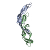 8enbSC 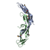 8endC S: Starting model for refinement C: citing same article ( |
|---|---|
| Similar structure data |
-Links
 PDBj
PDBj

- Assembly
| Deposited unit | 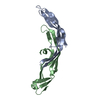
| ||||||||||||
|---|---|---|---|---|---|---|---|---|---|---|---|---|---|
| 1 |
| ||||||||||||
| Unit cell |
|
-Components
| #1: Protein | Mass: 10565.464 Da / Num. of mol.: 1 Source method: isolated from a genetically manipulated source Source: (gene. exp.)  Homo sapiens (human) / References: UniProt: A0T3A2 Homo sapiens (human) / References: UniProt: A0T3A2 |
|---|---|
| #2: Protein | Mass: 11590.098 Da / Num. of mol.: 1 Source method: isolated from a genetically manipulated source Source: (gene. exp.) Homo sapiens (human) / References: UniProt: A7DT38 |
| Has protein modification | Y |
-Experimental details
-Experiment
| Experiment | Method: X-RAY DIFFRACTION / Number of used crystals: 1 |
|---|
- Sample preparation
| Crystal | Density Matthews: 5.23 Å3/Da / Density % sol: 76.47 % |
|---|---|
| Crystal grow | Temperature: 293 K / Method: vapor diffusion, hanging drop Details: 0.2 M magnesium chloride, 0.1 M MES, pH 6.0, 50% w/v PEG200 |
-Data collection
| Diffraction | Mean temperature: 100 K / Serial crystal experiment: N |
|---|---|
| Diffraction source | Source: SYNCHROTRON / Site: APS / Beamline: 24-ID-E / Wavelength: 0.9792 Å |
| Detector | Type: ADSC QUANTUM 315 / Detector: CCD / Date: Oct 24, 2019 |
| Radiation | Protocol: SINGLE WAVELENGTH / Monochromatic (M) / Laue (L): M / Scattering type: x-ray |
| Radiation wavelength | Wavelength: 0.9792 Å / Relative weight: 1 |
| Reflection | Resolution: 3.29→44.87 Å / Num. obs: 6059 / % possible obs: 93.2 % / Redundancy: 19.7 % / Biso Wilson estimate: 165.38 Å2 / CC1/2: 0.999 / Rmerge(I) obs: 0.112 / Net I/σ(I): 14.1 |
| Reflection shell | Resolution: 3.29→3.57 Å / Num. unique obs: 404 / CC1/2: 0.389 |
- Processing
| Software |
| |||||||||||||||||||||||||||||||||||
|---|---|---|---|---|---|---|---|---|---|---|---|---|---|---|---|---|---|---|---|---|---|---|---|---|---|---|---|---|---|---|---|---|---|---|---|---|
| Refinement | Method to determine structure: MOLECULAR REPLACEMENT Starting model: PDB entry 8ENB Resolution: 3.29→44.87 Å / SU ML: 0.5182 / Cross valid method: FREE R-VALUE / σ(F): 1.34 / Phase error: 44.9477 Stereochemistry target values: GeoStd + Monomer Library + CDL v1.2
| |||||||||||||||||||||||||||||||||||
| Solvent computation | Shrinkage radii: 0.9 Å / VDW probe radii: 1.11 Å / Solvent model: FLAT BULK SOLVENT MODEL | |||||||||||||||||||||||||||||||||||
| Displacement parameters | Biso mean: 158.84 Å2 | |||||||||||||||||||||||||||||||||||
| Refinement step | Cycle: LAST / Resolution: 3.29→44.87 Å
| |||||||||||||||||||||||||||||||||||
| Refine LS restraints |
| |||||||||||||||||||||||||||||||||||
| LS refinement shell |
|
