 Movie
Movie Controller

[English] 日本語
 Yorodumi
- PDB-8elh: Crystal Structure of HLA-B*15:01 in complex with spike derived pe... -
+ Open data

- Basic information
| Entry | Database: PDB / ID: 8elh | |||||||||
|---|---|---|---|---|---|---|---|---|---|---|
| Title | Crystal Structure of HLA-B*15:01 in complex with spike derived peptide NQKLIANQF from SARS-CoV-2 virus | |||||||||
 Components Components |
| |||||||||
 Keywords Keywords | VIRAL PROTEIN/IMMUNE SYSTEM / TCR / T cell / HLA-B*15:01 / NQK / spike / SARS-CoV-2 / COVID / immune response / VIRAL PROTEIN-IMMUNE SYSTEM complex | |||||||||
| Function / homology |  Function and homology information Function and homology informationearly endosome lumen / Nef mediated downregulation of MHC class I complex cell surface expression / DAP12 interactions / Endosomal/Vacuolar pathway / Antigen Presentation: Folding, assembly and peptide loading of class I MHC / negative regulation of iron ion transport / T cell mediated cytotoxicity / cellular response to iron(III) ion / negative regulation of forebrain neuron differentiation / antigen processing and presentation of exogenous protein antigen via MHC class Ib, TAP-dependent ...early endosome lumen / Nef mediated downregulation of MHC class I complex cell surface expression / DAP12 interactions / Endosomal/Vacuolar pathway / Antigen Presentation: Folding, assembly and peptide loading of class I MHC / negative regulation of iron ion transport / T cell mediated cytotoxicity / cellular response to iron(III) ion / negative regulation of forebrain neuron differentiation / antigen processing and presentation of exogenous protein antigen via MHC class Ib, TAP-dependent / ER to Golgi transport vesicle membrane / peptide antigen assembly with MHC class I protein complex / transferrin transport / regulation of iron ion transport / regulation of erythrocyte differentiation / negative regulation of receptor-mediated endocytosis / HFE-transferrin receptor complex / response to molecule of bacterial origin / MHC class I peptide loading complex / cellular response to iron ion / positive regulation of T cell cytokine production / antigen processing and presentation of endogenous peptide antigen via MHC class I / MHC class I protein complex / peptide antigen assembly with MHC class II protein complex / negative regulation of neurogenesis / positive regulation of receptor-mediated endocytosis / cellular response to nicotine / MHC class II protein complex / positive regulation of T cell mediated cytotoxicity / multicellular organismal-level iron ion homeostasis / specific granule lumen / peptide antigen binding / antigen processing and presentation of exogenous peptide antigen via MHC class II / phagocytic vesicle membrane / positive regulation of immune response / recycling endosome membrane / Interferon gamma signaling / positive regulation of T cell activation / Immunoregulatory interactions between a Lymphoid and a non-Lymphoid cell / negative regulation of epithelial cell proliferation / Modulation by Mtb of host immune system / sensory perception of smell / positive regulation of cellular senescence / tertiary granule lumen / DAP12 signaling / MHC class II protein complex binding / T cell differentiation in thymus / late endosome membrane / negative regulation of neuron projection development / ER-Phagosome pathway / protein refolding / early endosome membrane / symbiont-mediated disruption of host tissue / Maturation of spike protein / Translation of Structural Proteins / Virion Assembly and Release / host cell surface / host extracellular space / symbiont-mediated-mediated suppression of host tetherin activity / amyloid fibril formation / Induction of Cell-Cell Fusion / structural constituent of virion / protein homotetramerization / intracellular iron ion homeostasis / membrane fusion / entry receptor-mediated virion attachment to host cell / Attachment and Entry / host cell endoplasmic reticulum-Golgi intermediate compartment membrane / positive regulation of viral entry into host cell / learning or memory / receptor-mediated virion attachment to host cell / host cell surface receptor binding / symbiont-mediated suppression of host innate immune response / endocytosis involved in viral entry into host cell / endoplasmic reticulum lumen / Amyloid fiber formation / receptor ligand activity / Golgi membrane / fusion of virus membrane with host plasma membrane / external side of plasma membrane / lysosomal membrane / focal adhesion / fusion of virus membrane with host endosome membrane / viral envelope / Neutrophil degranulation / symbiont entry into host cell / virion attachment to host cell / SARS-CoV-2 activates/modulates innate and adaptive immune responses / host cell plasma membrane / virion membrane / structural molecule activity / endoplasmic reticulum / Golgi apparatus / protein homodimerization activity / extracellular space / extracellular exosome / extracellular region / identical protein binding / membrane / plasma membrane Similarity search - Function | |||||||||
| Biological species |  Homo sapiens (human) Homo sapiens (human)  Severe acute respiratory syndrome coronavirus 2 Severe acute respiratory syndrome coronavirus 2 | |||||||||
| Method |  X-RAY DIFFRACTION / SYNCHROTRON / MOLECULAR REPLACEMENT / molecular replacement / Resolution: 1.85 Å X-RAY DIFFRACTION / SYNCHROTRON / MOLECULAR REPLACEMENT / molecular replacement / Resolution: 1.85 Å | |||||||||
 Authors Authors | Murdolo, L.D. / Gras, S. | |||||||||
| Funding support |  Australia, 2items Australia, 2items
| |||||||||
 Citation Citation | Journal: Nature / Year: 2023 Title: A common allele of HLA is associated with asymptomatic SARS-CoV-2 infection. Authors: Augusto, D.G. / Murdolo, L.D. / Chatzileontiadou, D.S.M. / Sabatino Jr., J.J. / Yusufali, T. / Peyser, N.D. / Butcher, X. / Kizer, K. / Guthrie, K. / Murray, V.W. / Pae, V. / ...Authors: Augusto, D.G. / Murdolo, L.D. / Chatzileontiadou, D.S.M. / Sabatino Jr., J.J. / Yusufali, T. / Peyser, N.D. / Butcher, X. / Kizer, K. / Guthrie, K. / Murray, V.W. / Pae, V. / Sarvadhavabhatla, S. / Beltran, F. / Gill, G.S. / Lynch, K.L. / Yun, C. / Maguire, C.T. / Peluso, M.J. / Hoh, R. / Henrich, T.J. / Deeks, S.G. / Davidson, M. / Lu, S. / Goldberg, S.A. / Kelly, J.D. / Martin, J.N. / Vierra-Green, C.A. / Spellman, S.R. / Langton, D.J. / Dewar-Oldis, M.J. / Smith, C. / Barnard, P.J. / Lee, S. / Marcus, G.M. / Olgin, J.E. / Pletcher, M.J. / Maiers, M. / Gras, S. / Hollenbach, J.A. | |||||||||
| History |
|
- Structure visualization
| Structure viewer | Molecule: MolmilJmol/JSmol |
|---|
- Downloads & links
-Download
| PDBx/mmCIF format | 8elh.cif.gz | 104.2 KB | Display | PDBx/mmCIF format |
|---|---|---|---|---|
| PDB format | pdb8elh.ent.gz | 77 KB | Display | PDB format |
| PDBx/mmJSON format | 8elh.json.gz | Tree view | PDBx/mmJSON format | |
| Others |  Other downloads Other downloads |
-Validation report
| Arichive directory | https://data.pdbj.org/pub/pdb/validation_reports/el/8elhftp://data.pdbj.org/pub/pdb/validation_reports/el/8elh | HTTPS FTP |
|---|
-Related structure data
| Related structure data |  8elgSC S: Starting model for refinement C: citing same article ( |
|---|---|
| Similar structure data |
-Links
 PDBj
PDBj


- Assembly
| Deposited unit | 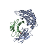
| ||||||||
|---|---|---|---|---|---|---|---|---|---|
| 1 |
| ||||||||
| Unit cell |
|
-Components
-Protein , 2 types, 2 molecules AB
| #1: Protein | Mass: 32132.352 Da / Num. of mol.: 1 Source method: isolated from a genetically manipulated source Source: (gene. exp.) Homo sapiens (human) / Plasmid: pET / Production host:  |
|---|---|
| #2: Protein | Mass: 11748.160 Da / Num. of mol.: 1 Source method: isolated from a genetically manipulated source Source: (gene. exp.) Homo sapiens (human) / Gene: B2M, CDABP0092, HDCMA22P / Plasmid: pET / Production host: |
-Protein/peptide , 1 types, 1 molecules C
| #3: Protein/peptide | Mass: 1076.225 Da / Num. of mol.: 1 / Fragment: NQK-OC43 peptide (UNP residues 919-927) / Source method: obtained synthetically Source: (synth.) Severe acute respiratory syndrome coronavirus 2References: UniProt: P0DTC2 |
|---|
-Non-polymers , 3 types, 331 molecules 


| #4: Chemical | ChemComp-ACT /  Mass: 59.044 Da / Num. of mol.: 1 / Source method: obtained synthetically / Formula: C2H3O2 Mass: 59.044 Da / Num. of mol.: 1 / Source method: obtained synthetically / Formula: C2H3O2 |
|---|---|
| #5: Chemical | ChemComp-PEG /  Mass: 106.120 Da / Num. of mol.: 1 / Source method: obtained synthetically / Formula: C4H10O3 Mass: 106.120 Da / Num. of mol.: 1 / Source method: obtained synthetically / Formula: C4H10O3 |
| #6: Water | ChemComp-HOH / Mass: 18.015 Da / Num. of mol.: 329 / Source method: isolated from a natural source / Formula: H2O |
-Details
| Has ligand of interest | N |
|---|---|
| Has protein modification | Y |
-Experimental details
-Experiment
| Experiment | Method: X-RAY DIFFRACTION / Number of used crystals: 1 |
|---|
- Sample preparation
| Crystal | Density Matthews: 2.53 Å3/Da / Density % sol: 51.32 % |
|---|---|
| Crystal grow | Temperature: 298 K / Method: vapor diffusion, hanging drop / pH: 8 / Details: 0.2 M sodium formate, pH 7.0, 20% w/v PEG3350 |
-Data collection
| Diffraction | Mean temperature: 100 K / Serial crystal experiment: N | ||||||||||||||||||||||||||||||
|---|---|---|---|---|---|---|---|---|---|---|---|---|---|---|---|---|---|---|---|---|---|---|---|---|---|---|---|---|---|---|---|
| Diffraction source | Source: SYNCHROTRON / Site: Australian Synchrotron / Beamline: MX2 / Wavelength: 0.956 Å | ||||||||||||||||||||||||||||||
| Detector | Type: DECTRIS PILATUS 6M / Detector: PIXEL / Date: Sep 21, 2022 | ||||||||||||||||||||||||||||||
| Radiation | Protocol: SINGLE WAVELENGTH / Monochromatic (M) / Laue (L): M / Scattering type: x-ray | ||||||||||||||||||||||||||||||
| Radiation wavelength | Wavelength: 0.956 Å / Relative weight: 1 | ||||||||||||||||||||||||||||||
| Reflection | Resolution: 1.85→45.56 Å / Num. obs: 39747 / % possible obs: 100 % / Redundancy: 13.5 % / CC1/2: 0.996 / Rmerge(I) obs: 0.217 / Rpim(I) all: 0.061 / Rrim(I) all: 0.226 / Net I/σ(I): 9.2 / Num. measured all: 535056 / Scaling rejects: 131 | ||||||||||||||||||||||||||||||
| Reflection shell | Diffraction-ID: 1
|
-Phasing
| Phasing | Method: molecular replacement |
|---|
- Processing
| Software |
| ||||||||||||||||||||||||||||||||||||||||||||||||||||||||||||||||||||||||||||||||||||||||||
|---|---|---|---|---|---|---|---|---|---|---|---|---|---|---|---|---|---|---|---|---|---|---|---|---|---|---|---|---|---|---|---|---|---|---|---|---|---|---|---|---|---|---|---|---|---|---|---|---|---|---|---|---|---|---|---|---|---|---|---|---|---|---|---|---|---|---|---|---|---|---|---|---|---|---|---|---|---|---|---|---|---|---|---|---|---|---|---|---|---|---|---|
| Refinement | Method to determine structure: MOLECULAR REPLACEMENT Starting model: PDB entry 8ELG Resolution: 1.85→43.08 Å / SU ML: 0.17 / Cross valid method: THROUGHOUT / σ(F): 1.34 / Phase error: 22.31 / Stereochemistry target values: ML
| ||||||||||||||||||||||||||||||||||||||||||||||||||||||||||||||||||||||||||||||||||||||||||
| Solvent computation | Shrinkage radii: 0.9 Å / VDW probe radii: 1.11 Å / Solvent model: FLAT BULK SOLVENT MODEL | ||||||||||||||||||||||||||||||||||||||||||||||||||||||||||||||||||||||||||||||||||||||||||
| Displacement parameters | Biso max: 86.36 Å2 / Biso mean: 27.3839 Å2 / Biso min: 10.31 Å2 | ||||||||||||||||||||||||||||||||||||||||||||||||||||||||||||||||||||||||||||||||||||||||||
| Refinement step | Cycle: final / Resolution: 1.85→43.08 Å
| ||||||||||||||||||||||||||||||||||||||||||||||||||||||||||||||||||||||||||||||||||||||||||
| LS refinement shell | Refine-ID: X-RAY DIFFRACTION / Rfactor Rfree error: 0 / Total num. of bins used: 14 / % reflection obs: 100 %
|
