 Movie
Movie Controller

[English] 日本語
 Yorodumi
- PDB-8ekt: CYP51 from Acanthamoeba castellanii in complex with the tetrazole... -
+ Open data

- Basic information
| Entry | Database: PDB / ID: 8ekt | ||||||
|---|---|---|---|---|---|---|---|
| Title | CYP51 from Acanthamoeba castellanii in complex with the tetrazole-based IND inhibitor VT-1161(VT1) | ||||||
 Components Components | sterol 14a-demethylase | ||||||
 Keywords Keywords | OXIDOREDUCTASE/INHIBITOR / cytochrome P450 / CYP51 / sterol 14alpha-demethylase / sterol biosynthesis / OXIDOREDUCTASE-INHIBITOR complex | ||||||
| Function / homology |  Function and homology information Function and homology informationoxidoreductase activity, acting on paired donors, with incorporation or reduction of molecular oxygen / methyltransferase activity / monooxygenase activity / methylation / iron ion binding / heme binding Similarity search - Function | ||||||
| Biological species |  Acanthamoeba castellanii (eukaryote) Acanthamoeba castellanii (eukaryote) | ||||||
| Method |  X-RAY DIFFRACTION / SYNCHROTRON / MOLECULAR REPLACEMENT / Resolution: 2.29 Å X-RAY DIFFRACTION / SYNCHROTRON / MOLECULAR REPLACEMENT / Resolution: 2.29 Å | ||||||
 Authors Authors | Hargrove, T.Y. / Wawrzak, Z. / Lepesheva, G.I. | ||||||
| Funding support |  United States, 1items United States, 1items
| ||||||
 Citation Citation | Journal: J.Med.Chem. / Year: 2024 Title: Identification of Potent and Selective Inhibitors of Acanthamoeba : Structural Insights into Sterol 14 alpha-Demethylase as a Key Drug Target. Authors: Hargrove, T.Y. / Lamb, D.C. / Wawrzak, Z. / Hull, M. / Kelly, S.L. / Guengerich, F.P. / Lepesheva, G.I. | ||||||
| History |
|
- Structure visualization
| Structure viewer | Molecule: MolmilJmol/JSmol |
|---|
- Downloads & links
-Download
| PDBx/mmCIF format | 8ekt.cif.gz | 515.7 KB | Display | PDBx/mmCIF format |
|---|---|---|---|---|
| PDB format | pdb8ekt.ent.gz | 423.9 KB | Display | PDB format |
| PDBx/mmJSON format | 8ekt.json.gz | Tree view | PDBx/mmJSON format | |
| Others |  Other downloads Other downloads |
-Validation report
| Arichive directory | https://data.pdbj.org/pub/pdb/validation_reports/ek/8ektftp://data.pdbj.org/pub/pdb/validation_reports/ek/8ekt | HTTPS FTP |
|---|
-Related structure data
| Related structure data |  7uwpS S: Starting model for refinement |
|---|---|
| Similar structure data |
-Links
 PDBj
PDBj
- Assembly
| Deposited unit | 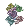
| ||||||||
|---|---|---|---|---|---|---|---|---|---|
| 1 | 
| ||||||||
| 2 | 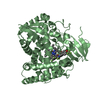
| ||||||||
| 3 | 
| ||||||||
| 4 | 
| ||||||||
| 5 | 
| ||||||||
| 6 | 
| ||||||||
| Unit cell |
|
-Components
| #1: Protein | Mass: 52447.324 Da / Num. of mol.: 6 Source method: isolated from a genetically manipulated source Source: (gene. exp.) Acanthamoeba castellanii (eukaryote) / Plasmid: pCW / Production host:  #2: Chemical | ChemComp-HEM /   Mass: 616.487 Da / Num. of mol.: 6 / Source method: obtained synthetically / Formula: C34H32FeN4O4 Mass: 616.487 Da / Num. of mol.: 6 / Source method: obtained synthetically / Formula: C34H32FeN4O4#3: Chemical | ChemComp-VT1 / ( 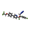  Mass: 527.394 Da / Num. of mol.: 6 / Source method: obtained synthetically / Formula: C23H16F7N5O2 / Feature type: SUBJECT OF INVESTIGATION Mass: 527.394 Da / Num. of mol.: 6 / Source method: obtained synthetically / Formula: C23H16F7N5O2 / Feature type: SUBJECT OF INVESTIGATION#4: Water | ChemComp-HOH / |  Mass: 18.015 Da / Num. of mol.: 448 / Source method: isolated from a natural source / Formula: H2O Mass: 18.015 Da / Num. of mol.: 448 / Source method: isolated from a natural source / Formula: H2OHas ligand of interest | Y | Has protein modification | N | |
|---|
-Experimental details
-Experiment
| Experiment | Method: X-RAY DIFFRACTION / Number of used crystals: 1 |
|---|
- Sample preparation
| Crystal | Density Matthews: 3.04 Å3/Da / Density % sol: 59 % |
|---|---|
| Crystal grow | Temperature: 298 K / Method: vapor diffusion, hanging drop / pH: 6.7 Details: 18% PEG8000, 0.1 M sodium citrate, 0.34 mM n-dodecyl-b-D-maltoside 5.5 mM TCEP |
-Data collection
| Diffraction | Mean temperature: 100 K / Serial crystal experiment: N |
|---|---|
| Diffraction source | Source: SYNCHROTRON / Site: APS / Beamline: 21-ID-G / Wavelength: 0.97857 Å |
| Detector | Type: RAYONIX MX-300 / Detector: CCD / Date: Sep 16, 2022 / Details: Be lens |
| Radiation | Monochromator: C(111) / Protocol: SINGLE WAVELENGTH / Monochromatic (M) / Laue (L): M / Scattering type: x-ray |
| Radiation wavelength | Wavelength: 0.97857 Å / Relative weight: 1 |
| Reflection | Resolution: 2.28→30 Å / Num. obs: 118963 / % possible obs: 98.3 % / Redundancy: 3.9 % / Biso Wilson estimate: 32.7 Å2 / CC1/2: 0.993 / Rmerge(I) obs: 0.04 / Net I/σ(I): 25 |
| Reflection shell | Resolution: 2.28→2.32 Å / Rmerge(I) obs: 1.02 / Mean I/σ(I) obs: 1.7 / Num. unique obs: 1585 / CC1/2: 0.539 |
- Processing
| Software |
| |||||||||||||||||||||||||||||||||||||||||||||||||||||||
|---|---|---|---|---|---|---|---|---|---|---|---|---|---|---|---|---|---|---|---|---|---|---|---|---|---|---|---|---|---|---|---|---|---|---|---|---|---|---|---|---|---|---|---|---|---|---|---|---|---|---|---|---|---|---|---|---|
| Refinement | Method to determine structure: MOLECULAR REPLACEMENT Starting model: PDB entry 7UWP Resolution: 2.29→29.89 Å / Cor.coef. Fo:Fc: 0.93 / Cor.coef. Fo:Fc free: 0.921 / SU B: 6.312 / SU ML: 0.148 / Cross valid method: THROUGHOUT / σ(F): 0 / ESU R: 0.414 / ESU R Free: 0.237 / Stereochemistry target values: MAXIMUM LIKELIHOOD Details: HYDROGENS HAVE BEEN ADDED IN THE RIDING POSITIONS U VALUES : REFINED INDIVIDUALLY
| |||||||||||||||||||||||||||||||||||||||||||||||||||||||
| Solvent computation | Ion probe radii: 0.8 Å / Shrinkage radii: 0.8 Å / VDW probe radii: 1.2 Å / Solvent model: MASK | |||||||||||||||||||||||||||||||||||||||||||||||||||||||
| Displacement parameters | Biso max: 155.93 Å2 / Biso mean: 44.581 Å2 / Biso min: 8.12 Å2
| |||||||||||||||||||||||||||||||||||||||||||||||||||||||
| Refinement step | Cycle: final / Resolution: 2.29→29.89 Å
| |||||||||||||||||||||||||||||||||||||||||||||||||||||||
| Refine LS restraints |
| |||||||||||||||||||||||||||||||||||||||||||||||||||||||
| LS refinement shell | Resolution: 2.292→2.351 Å / Rfactor Rfree error: 0 / Total num. of bins used: 20
|
