 Movie
Movie Controller


+ Open data

- Basic information
| Entry | Database: PDB / ID: 8eko | ||||||
|---|---|---|---|---|---|---|---|
| Title | Sperm whale myoglobin mutant L29H F33W F43H (F33W CuBMb) | ||||||
 Components Components | Myoglobin | ||||||
 Keywords Keywords | OXIDOREDUCTASE / Oxygen reduction reaction / heme-copper oxidase / biosynthetic model | ||||||
| Function / homology |  Function and homology information Function and homology informationOxidoreductases; Acting on other nitrogenous compounds as donors / nitrite reductase activity / sarcoplasm / Oxidoreductases; Acting on a peroxide as acceptor; Peroxidases / removal of superoxide radicals / oxygen carrier activity / peroxidase activity / oxygen binding / heme binding / extracellular exosome / metal ion binding Similarity search - Function | ||||||
| Biological species |  | ||||||
| Method |  X-RAY DIFFRACTION / SYNCHROTRON / MOLECULAR REPLACEMENT / Resolution: 1.34 Å X-RAY DIFFRACTION / SYNCHROTRON / MOLECULAR REPLACEMENT / Resolution: 1.34 Å | ||||||
 Authors Authors | Ledray, A.P. / Dwaraknath, S. / Lu, Y. | ||||||
| Funding support |  United States, 1items United States, 1items
| ||||||
 Citation Citation | Journal: Biochemistry / Year: 2023 Title: Tryptophan Can Promote Oxygen Reduction to Water in a Biosynthetic Model of Heme Copper Oxidases. Authors: Ledray, A.P. / Dwaraknath, S. / Chakarawet, K. / Sponholtz, M.R. / Merchen, C. / Van Stappen, C. / Rao, G. / Britt, R.D. / Lu, Y. | ||||||
| History |
|
- Structure visualization
| Structure viewer | Molecule: MolmilJmol/JSmol |
|---|
- Downloads & links
-Download
| PDBx/mmCIF format | 8eko.cif.gz | 107.5 KB | Display | PDBx/mmCIF format |
|---|---|---|---|---|
| PDB format | pdb8eko.ent.gz | 67.4 KB | Display | PDB format |
| PDBx/mmJSON format | 8eko.json.gz | Tree view | PDBx/mmJSON format | |
| Others |  Other downloads Other downloads |
-Validation report
| Summary document | 8eko_validation.pdf.gz | 1.1 MB | Display | wwPDB validaton report |
|---|---|---|---|---|
| Full document | 8eko_full_validation.pdf.gz | 1.1 MB | Display | |
| Data in XML | 8eko_validation.xml.gz | 10 KB | Display | |
| Data in CIF | 8eko_validation.cif.gz | 14 KB | Display | |
| Arichive directory | https://data.pdbj.org/pub/pdb/validation_reports/ek/8ekoftp://data.pdbj.org/pub/pdb/validation_reports/ek/8eko | HTTPS FTP |
-Related structure data
| Related structure data | 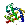 4fwyS S: Starting model for refinement |
|---|---|
| Similar structure data |
-Links
 PDBj
PDBj


- Assembly
| Deposited unit | 
| ||||||||||||
|---|---|---|---|---|---|---|---|---|---|---|---|---|---|
| 1 |
| ||||||||||||
| Unit cell |
|
-Components
| #1: Protein | Mass: 17289.951 Da / Num. of mol.: 1 / Mutation: L29H, F33W, F43H Source method: isolated from a genetically manipulated source Source: (gene. exp.)  |
|---|---|
| #2: Chemical | ChemComp-HEM /   Mass: 616.487 Da / Num. of mol.: 1 / Source method: obtained synthetically / Formula: C34H32FeN4O4 / Feature type: SUBJECT OF INVESTIGATION Mass: 616.487 Da / Num. of mol.: 1 / Source method: obtained synthetically / Formula: C34H32FeN4O4 / Feature type: SUBJECT OF INVESTIGATION |
| #3: Water | ChemComp-HOH /  Mass: 18.015 Da / Num. of mol.: 139 / Source method: isolated from a natural source / Formula: H2O Mass: 18.015 Da / Num. of mol.: 139 / Source method: isolated from a natural source / Formula: H2O |
| Has ligand of interest | Y |
-Experimental details
-Experiment
| Experiment | Method: X-RAY DIFFRACTION / Number of used crystals: 1 |
|---|
- Sample preparation
| Crystal | Density Matthews: 2.16 Å3/Da / Density % sol: 43.07 % |
|---|---|
| Crystal grow | Temperature: 277 K / Method: vapor diffusion, hanging drop / pH: 6.5 Details: F33W CuBMb (1.0 mM) in 100 mM tris pH 8, (pH adjusted H2SO4), was mixed 1:3 with well buffer (0.1 M sodium cacodylate pH 6.5, 0.2 M sodium acetate trihydrate with 30% w/v polyethylene glycol ...Details: F33W CuBMb (1.0 mM) in 100 mM tris pH 8, (pH adjusted H2SO4), was mixed 1:3 with well buffer (0.1 M sodium cacodylate pH 6.5, 0.2 M sodium acetate trihydrate with 30% w/v polyethylene glycol 8000) using the hanging drop method with 300 uL well buffer in the well of the crystallization tray. Crystals obtained by this method were soaked with 10 eq CuSO4 in a mixture of F33W CuBMb and well buffer consistent with the original drop conditions to avoid dissolving the crystals and subsequently with 15eq potassium cyanide, in a mixture of E-F33W-CuBMb and well buffer using pH 7.0 sodium cacodylate |
-Data collection
| Diffraction | Mean temperature: 100 K / Serial crystal experiment: N |
|---|---|
| Diffraction source | Source: SYNCHROTRON / Site: ALS / Beamline: 5.0.2 / Wavelength: 1 Å |
| Detector | Type: DECTRIS PILATUS3 6M / Detector: PIXEL / Date: Jun 14, 2019 |
| Radiation | Monochromator: Double-crystal, Si(111) / Protocol: SINGLE WAVELENGTH / Monochromatic (M) / Laue (L): M / Scattering type: x-ray |
| Radiation wavelength | Wavelength: 1 Å / Relative weight: 1 |
| Reflection | Resolution: 1.34→35.61 Å / Num. obs: 34356 / % possible obs: 99.91 % / Redundancy: 2 % / Biso Wilson estimate: 9.35 Å2 / CC1/2: 0.999 / CC star: 1 / Rmerge(I) obs: 0.1921 / Rrim(I) all: 0.02717 / Net I/σ(I): 19.35 |
| Reflection shell | Resolution: 1.34→1.39 Å / Redundancy: 2 % / Rmerge(I) obs: 0.05665 / Num. unique obs: 3381 / CC1/2: 0.987 / CC star: 0.997 / Rrim(I) all: 0.08011 / % possible all: 99.71 |
- Processing
| Software |
| |||||||||||||||||||||||||||||||||||||||||||||||||||||||||||||||||||||||||||||||||||||||||||
|---|---|---|---|---|---|---|---|---|---|---|---|---|---|---|---|---|---|---|---|---|---|---|---|---|---|---|---|---|---|---|---|---|---|---|---|---|---|---|---|---|---|---|---|---|---|---|---|---|---|---|---|---|---|---|---|---|---|---|---|---|---|---|---|---|---|---|---|---|---|---|---|---|---|---|---|---|---|---|---|---|---|---|---|---|---|---|---|---|---|---|---|---|
| Refinement | Method to determine structure: MOLECULAR REPLACEMENT Starting model: 4FWY Resolution: 1.34→35.61 Å / SU ML: 0.0795 / Cross valid method: FREE R-VALUE / σ(F): 1.35 / Phase error: 13.9685 Stereochemistry target values: GeoStd + Monomer Library + CDL v1.2
| |||||||||||||||||||||||||||||||||||||||||||||||||||||||||||||||||||||||||||||||||||||||||||
| Solvent computation | Shrinkage radii: 0.9 Å / VDW probe radii: 1.11 Å / Solvent model: FLAT BULK SOLVENT MODEL | |||||||||||||||||||||||||||||||||||||||||||||||||||||||||||||||||||||||||||||||||||||||||||
| Displacement parameters | Biso mean: 15.53 Å2 | |||||||||||||||||||||||||||||||||||||||||||||||||||||||||||||||||||||||||||||||||||||||||||
| Refinement step | Cycle: LAST / Resolution: 1.34→35.61 Å
| |||||||||||||||||||||||||||||||||||||||||||||||||||||||||||||||||||||||||||||||||||||||||||
| Refine LS restraints |
| |||||||||||||||||||||||||||||||||||||||||||||||||||||||||||||||||||||||||||||||||||||||||||
| LS refinement shell |
|
