 Movie
Movie Controller


+ Open data

- Basic information
| Entry | Database: PDB / ID: 8eke | ||||||
|---|---|---|---|---|---|---|---|
| Title | Cryo-EM structure of SARS CoV-2 Mpro WT protease | ||||||
 Components Components | 3C-like proteinase nsp5 | ||||||
 Keywords Keywords | VIRAL PROTEIN / Main protease / Mpro / SARS CoV-2 / polyprotein | ||||||
| Function / homology |  Function and homology information Function and homology informationprotein guanylyltransferase activity / RNA endonuclease activity producing 3'-phosphomonoesters, hydrolytic mechanism / 5'-3' RNA helicase activity / Lyases; Phosphorus-oxygen lyases / Assembly of the SARS-CoV-2 Replication-Transcription Complex (RTC) / symbiont-mediated suppression of host cytoplasmic pattern recognition receptor signaling pathway via inhibition of TBK1 activity / Maturation of replicase proteins / TRAF3-dependent IRF activation pathway / ISG15-specific peptidase activity / Transcription of SARS-CoV-2 sgRNAs ...protein guanylyltransferase activity / RNA endonuclease activity producing 3'-phosphomonoesters, hydrolytic mechanism / 5'-3' RNA helicase activity / Lyases; Phosphorus-oxygen lyases / Assembly of the SARS-CoV-2 Replication-Transcription Complex (RTC) / symbiont-mediated suppression of host cytoplasmic pattern recognition receptor signaling pathway via inhibition of TBK1 activity / Maturation of replicase proteins / TRAF3-dependent IRF activation pathway / ISG15-specific peptidase activity / Transcription of SARS-CoV-2 sgRNAs / snRNP Assembly / Translation of Replicase and Assembly of the Replication Transcription Complex / Replication of the SARS-CoV-2 genome / Hydrolases; Acting on ester bonds; Exoribonucleases producing 5'-phosphomonoesters / double membrane vesicle viral factory outer membrane / SARS coronavirus main proteinase / host cell endoplasmic reticulum-Golgi intermediate compartment / host cell endosome / 3'-5'-RNA exonuclease activity / symbiont-mediated degradation of host mRNA / 5'-3' DNA helicase activity / mRNA guanylyltransferase / symbiont-mediated suppression of host toll-like receptor signaling pathway / symbiont-mediated suppression of host ISG15-protein conjugation / G-quadruplex RNA binding / mRNA guanylyltransferase activity / symbiont-mediated suppression of host cytoplasmic pattern recognition receptor signaling pathway via inhibition of IRF3 activity / DNA helicase / omega peptidase activity / mRNA (guanine-N7)-methyltransferase / methyltransferase cap1 / symbiont-mediated suppression of host NF-kappaB cascade / SARS-CoV-2 modulates host translation machinery / symbiont-mediated perturbation of host ubiquitin-like protein modification / host cell Golgi apparatus / methyltransferase cap1 activity / mRNA 5'-cap (guanine-N7-)-methyltransferase activity / ubiquitinyl hydrolase 1 / Hydrolases; Acting on peptide bonds (peptidases); Cysteine endopeptidases / lyase activity / cysteine-type deubiquitinase activity / single-stranded RNA binding / viral protein processing / host cell perinuclear region of cytoplasm / host cell endoplasmic reticulum membrane / RNA helicase / symbiont-mediated suppression of host type I interferon-mediated signaling pathway / symbiont-mediated suppression of host gene expression / copper ion binding / viral translational frameshifting / symbiont-mediated activation of host autophagy / RNA-directed RNA polymerase / cysteine-type endopeptidase activity / viral RNA genome replication / RNA-directed RNA polymerase activity / lipid binding / host cell nucleus / SARS-CoV-2 activates/modulates innate and adaptive immune responses / ATP hydrolysis activity / DNA-templated transcription / proteolysis / RNA binding / zinc ion binding / ATP binding Similarity search - Function | ||||||
| Biological species |   Severe acute respiratory syndrome coronavirus 2 Severe acute respiratory syndrome coronavirus 2 | ||||||
| Method | ELECTRON MICROSCOPY / single particle reconstruction / cryo EM / Resolution: 3.36 Å | ||||||
 Authors Authors | Narwal, M. / Edwards, T. / Armache, J.P. / Murakami, K.S. | ||||||
| Funding support |  United States, 1items United States, 1items
| ||||||
 Citation Citation | Journal: J Biol Chem / Year: 2023 Title: SARS-CoV-2 polyprotein substrate regulates the stepwise M cleavage reaction. Authors: Manju Narwal / Jean-Paul Armache / Thomas J Edwards / Katsuhiko S Murakami / Abstract: The processing of the Coronavirus polyproteins pp1a and pp1ab by the main protease M to produce mature proteins is a crucial event in virus replication and a promising target for antiviral drug ...The processing of the Coronavirus polyproteins pp1a and pp1ab by the main protease M to produce mature proteins is a crucial event in virus replication and a promising target for antiviral drug development. M cleaves polyproteins in a defined order, but how M and/or the polyproteins determine the order of cleavage remains enigmatic due to a lack of structural information about polyprotein-bound M. Here, we present the cryo-EM structures of SARS-CoV-2 M in an apo form and in complex with the nsp7-10 region of the pp1a polyprotein. The complex structure shows that M interacts with only the recognition site residues between nsp9 and nsp10, without any association with the rest of the polyprotein. Comparison between the apo form and polyprotein-bound structures of M highlights the flexible nature of the active site region of M, which allows it to accommodate ten recognition sites found in the polyprotein. These observations suggest that the role of M in selecting a preferred cleavage site is limited and underscores the roles of the structure, conformation, and/or dynamics of the polyproteins in determining the sequence of polyprotein cleavage by M. | ||||||
| History |
|
- Structure visualization
| Structure viewer | Molecule: MolmilJmol/JSmol |
|---|
- Downloads & links
-Download
| PDBx/mmCIF format | 8eke.cif.gz | 150.7 KB | Display | PDBx/mmCIF format |
|---|---|---|---|---|
| PDB format | pdb8eke.ent.gz | 95.5 KB | Display | PDB format |
| PDBx/mmJSON format | 8eke.json.gz | Tree view | PDBx/mmJSON format | |
| Others |  Other downloads Other downloads |
-Validation report
| Arichive directory | https://data.pdbj.org/pub/pdb/validation_reports/ek/8ekeftp://data.pdbj.org/pub/pdb/validation_reports/ek/8eke | HTTPS FTP |
|---|
-Related structure data
| Related structure data |  28200MC 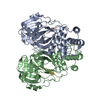 8eirC M: map data used to model this data C: citing same article ( |
|---|---|
| Similar structure data |

-Links
 PDBj
PDBj


- Assembly
| Deposited unit | 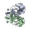
|
|---|---|
| 1 |
|
-Components
| #1: Protein | Mass: 33793.480 Da / Num. of mol.: 2 / Mutation: C145A Source method: isolated from a genetically manipulated source Source: (gene. exp.) Severe acute respiratory syndrome coronavirus 2Gene: rep, 1a-1b Production host:  References: UniProt: P0DTD1, SARS coronavirus main proteinase Compound details | Main protease (Mpro) of SARS CoV-2 virus. The protease is critical for the processing of pp1a/b ...Main protease (Mpro) of SARS CoV-2 virus. The protease is critical for the processing of pp1a/b polyprotein for the release of functional non-structural proteins. | |
|---|
-Experimental details
-Experiment
| Experiment | Method: ELECTRON MICROSCOPY |
|---|---|
| EM experiment | Aggregation state: PARTICLE / 3D reconstruction method: single particle reconstruction |
- Sample preparation
| Component | Name: SARS CoV-2 Main protease (Mpro) nsp5 apo structure / Type: COMPLEX / Entity ID: all / Source: RECOMBINANT | ||||||||||||||||||||
|---|---|---|---|---|---|---|---|---|---|---|---|---|---|---|---|---|---|---|---|---|---|
| Molecular weight | Value: 0.07 MDa / Experimental value: YES | ||||||||||||||||||||
| Source (natural) | Organism: Severe acute respiratory syndrome coronavirus 2 | ||||||||||||||||||||
| Source (recombinant) | Organism: | ||||||||||||||||||||
| Buffer solution | pH: 7.5 | ||||||||||||||||||||
| Buffer component |
| ||||||||||||||||||||
| Specimen | Conc.: 1 mg/ml / Embedding applied: NO / Shadowing applied: NO / Staining applied: NO / Vitrification applied: YES / Details: Homogenous sample | ||||||||||||||||||||
| Vitrification | Instrument: FEI VITROBOT MARK IV / Cryogen name: ETHANE / Humidity: 100 % / Chamber temperature: 277 K |
- Electron microscopy imaging
| Experimental equipment |  Model: Titan Krios / Image courtesy: FEI Company |
|---|---|
| Microscopy | Model: FEI TITAN KRIOS |
| Electron gun | Electron source:  FIELD EMISSION GUN / Accelerating voltage: 300 kV / Illumination mode: SPOT SCAN FIELD EMISSION GUN / Accelerating voltage: 300 kV / Illumination mode: SPOT SCAN |
| Electron lens | Mode: OTHER / Nominal defocus max: 2000 nm / Nominal defocus min: 750 nm |
| Image recording | Electron dose: 50 e/Å2 / Film or detector model: GATAN K3 (6k x 4k) |
- Processing
| Software |
| ||||||||||||||||||||||||
|---|---|---|---|---|---|---|---|---|---|---|---|---|---|---|---|---|---|---|---|---|---|---|---|---|---|
| CTF correction | Type: PHASE FLIPPING AND AMPLITUDE CORRECTION | ||||||||||||||||||||||||
| 3D reconstruction | Resolution: 3.36 Å / Resolution method: FSC 0.143 CUT-OFF / Num. of particles: 95597 / Symmetry type: POINT | ||||||||||||||||||||||||
| Refinement | Cross valid method: NONE Stereochemistry target values: GeoStd + Monomer Library + CDL v1.2 | ||||||||||||||||||||||||
| Displacement parameters | Biso mean: 94.74 Å2 | ||||||||||||||||||||||||
| Refine LS restraints |
|
