 Movie
Movie Controller

[English] 日本語
 Yorodumi
- PDB-8ek7: Crystal structure of Hepes and Mg bound 2,3-diketo-5-methylthiope... -
+ Open data

- Basic information
| Entry | Database: PDB / ID: 8ek7 | |||||||||
|---|---|---|---|---|---|---|---|---|---|---|
| Title | Crystal structure of Hepes and Mg bound 2,3-diketo-5-methylthiopentyl-1-phosphate enolase-phosphatase from Klebsiella aerogenes | |||||||||
 Components Components | Orotidine 5'-phosphate decarboxylase | |||||||||
 Keywords Keywords | ISOMERASE / SSGCID / STRUCTURAL GENOMICS / SEATTLE STRUCTURAL GENOMICS CENTER FOR INFECTIOUS DISEASE | |||||||||
| Function / homology |  Function and homology information Function and homology informationorotidine-5'-phosphate decarboxylase / orotidine-5'-phosphate decarboxylase activity / 'de novo' UMP biosynthetic process / 'de novo' pyrimidine nucleobase biosynthetic process Similarity search - Function | |||||||||
| Biological species |  Klebsiella aerogenes (bacteria) Klebsiella aerogenes (bacteria) | |||||||||
| Method |  X-RAY DIFFRACTION / SYNCHROTRON / MOLECULAR REPLACEMENT / molecular replacement / Resolution: 1.65 Å X-RAY DIFFRACTION / SYNCHROTRON / MOLECULAR REPLACEMENT / molecular replacement / Resolution: 1.65 Å | |||||||||
 Authors Authors | Seattle Structural Genomics Center for Infectious Disease / Seattle Structural Genomics Center for Infectious Disease (SSGCID) | |||||||||
| Funding support |  United States, 2items United States, 2items
| |||||||||
 Citation Citation | Journal: To be published Title: Crystal structure of Hepes and Mg bound 2,3-diketo-5-methylthiopentyl-1-phosphate enolase-phosphatase from Klebsiella aerogenes Authors: Lovell, S. / Liu, L. / Seibold, S. / Battaile, K.P. | |||||||||
| History |
|
- Structure visualization
| Structure viewer | Molecule: MolmilJmol/JSmol |
|---|
- Downloads & links
-Download
| PDBx/mmCIF format | 8ek7.cif.gz | 274.9 KB | Display | PDBx/mmCIF format |
|---|---|---|---|---|
| PDB format | pdb8ek7.ent.gz | 219.2 KB | Display | PDB format |
| PDBx/mmJSON format | 8ek7.json.gz | Tree view | PDBx/mmJSON format | |
| Others |  Other downloads Other downloads |
-Validation report
| Arichive directory | https://data.pdbj.org/pub/pdb/validation_reports/ek/8ek7ftp://data.pdbj.org/pub/pdb/validation_reports/ek/8ek7 | HTTPS FTP |
|---|
-Related structure data
| Related structure data |  8dopS S: Starting model for refinement |
|---|---|
| Similar structure data |
-Links
 PDBj
PDBj
- Assembly
| Deposited unit | 
| ||||||||
|---|---|---|---|---|---|---|---|---|---|
| 1 | 
| ||||||||
| 2 | 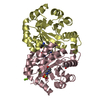
| ||||||||
| 3 | 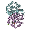
| ||||||||
| Unit cell |
|
-Components
| #1: Protein | Mass: 27348.453 Da / Num. of mol.: 6 Source method: isolated from a genetically manipulated source Source: (gene. exp.) Klebsiella aerogenes (bacteria) / Gene: pyrF, BXQ27_18035, HV316_11080 / Plasmid: KlaeA.01229.a.B1 / Production host: References: UniProt: A0A0M3H420, orotidine-5'-phosphate decarboxylase #2: Chemical |   Mass: 238.305 Da / Num. of mol.: 3 / Source method: obtained synthetically / Formula: C8H18N2O4S / Comment: pH buffer*YM Mass: 238.305 Da / Num. of mol.: 3 / Source method: obtained synthetically / Formula: C8H18N2O4S / Comment: pH buffer*YM#3: Chemical | ChemComp-MG /   Mass: 24.305 Da / Num. of mol.: 10 / Source method: obtained synthetically / Formula: Mg Mass: 24.305 Da / Num. of mol.: 10 / Source method: obtained synthetically / Formula: Mg#4: Chemical | ChemComp-MPD / (   Mass: 118.174 Da / Num. of mol.: 5 / Source method: obtained synthetically / Formula: C6H14O2 / Comment: precipitant*YM Mass: 118.174 Da / Num. of mol.: 5 / Source method: obtained synthetically / Formula: C6H14O2 / Comment: precipitant*YM#5: Water | ChemComp-HOH / |  Mass: 18.015 Da / Num. of mol.: 685 / Source method: isolated from a natural source / Formula: H2O Mass: 18.015 Da / Num. of mol.: 685 / Source method: isolated from a natural source / Formula: H2OHas ligand of interest | N | |
|---|
-Experimental details
-Experiment
| Experiment | Method: X-RAY DIFFRACTION / Number of used crystals: 1 |
|---|
- Sample preparation
| Crystal | Density Matthews: 2.07 Å3/Da / Density % sol: 40.52 % |
|---|---|
| Crystal grow | Temperature: 291 K / Method: vapor diffusion, sitting drop / pH: 7.5 Details: Morpheus A8: 12.5%(v/v) MPD, 12.5%(v/v) PEG 1000, 12.5%(w/v) PEG 3350, 100 mM HEPES/MOPS, pH 7.5, 30 mM MgCl2 and 30 mM CaCl2, KlaeA.01229.a.B1.PW39096 at 15 mg/mL. plate 12527, well A8 drop ...Details: Morpheus A8: 12.5%(v/v) MPD, 12.5%(v/v) PEG 1000, 12.5%(w/v) PEG 3350, 100 mM HEPES/MOPS, pH 7.5, 30 mM MgCl2 and 30 mM CaCl2, KlaeA.01229.a.B1.PW39096 at 15 mg/mL. plate 12527, well A8 drop 3, Puck: PSL0308, Cryo: Direct |
-Data collection
| Diffraction | Mean temperature: 100 K / Serial crystal experiment: N | |||||||||||||||||||||
|---|---|---|---|---|---|---|---|---|---|---|---|---|---|---|---|---|---|---|---|---|---|---|
| Diffraction source | Source: SYNCHROTRON / Site: NSLS-II / Beamline: 19-ID / Wavelength: 0.97856 Å | |||||||||||||||||||||
| Detector | Type: DECTRIS EIGER2 XE 9M / Detector: PIXEL / Date: Aug 11, 2022 | |||||||||||||||||||||
| Radiation | Monochromator: Double Crystal Si 111 / Protocol: SINGLE WAVELENGTH / Monochromatic (M) / Laue (L): M / Scattering type: x-ray | |||||||||||||||||||||
| Radiation wavelength | Wavelength: 0.97856 Å / Relative weight: 1 | |||||||||||||||||||||
| Reflection | Resolution: 1.65→49.41 Å / Num. obs: 160347 / % possible obs: 99.7 % / Redundancy: 3.6 % / Biso Wilson estimate: 18.9 Å2 / CC1/2: 0.999 / Rmerge(I) obs: 0.048 / Net I/σ(I): 11.5 / Num. measured all: 581276 / Scaling rejects: 7 | |||||||||||||||||||||
| Reflection shell | Diffraction-ID: 1 / Redundancy: 3.5 %
|
-Phasing
| Phasing | Method: molecular replacement |
|---|
- Processing
| Software |
| |||||||||||||||||||||||||||||||||||||||||||||||||||||||||||||||||||||||||||||||||||||||||||||||||||||||||||||||||||||||||||||||||||||||||||||||||||||||||||||||||||||||||||||||||||||||||||||||||||||||||||||||||||||||||
|---|---|---|---|---|---|---|---|---|---|---|---|---|---|---|---|---|---|---|---|---|---|---|---|---|---|---|---|---|---|---|---|---|---|---|---|---|---|---|---|---|---|---|---|---|---|---|---|---|---|---|---|---|---|---|---|---|---|---|---|---|---|---|---|---|---|---|---|---|---|---|---|---|---|---|---|---|---|---|---|---|---|---|---|---|---|---|---|---|---|---|---|---|---|---|---|---|---|---|---|---|---|---|---|---|---|---|---|---|---|---|---|---|---|---|---|---|---|---|---|---|---|---|---|---|---|---|---|---|---|---|---|---|---|---|---|---|---|---|---|---|---|---|---|---|---|---|---|---|---|---|---|---|---|---|---|---|---|---|---|---|---|---|---|---|---|---|---|---|---|---|---|---|---|---|---|---|---|---|---|---|---|---|---|---|---|---|---|---|---|---|---|---|---|---|---|---|---|---|---|---|---|---|---|---|---|---|---|---|---|---|---|---|---|---|---|---|---|---|
| Refinement | Method to determine structure: MOLECULAR REPLACEMENT Starting model: 8DOP Resolution: 1.65→41.69 Å / SU ML: 0.19 / Cross valid method: THROUGHOUT / σ(F): 1 / Phase error: 28.67 / Stereochemistry target values: ML
| |||||||||||||||||||||||||||||||||||||||||||||||||||||||||||||||||||||||||||||||||||||||||||||||||||||||||||||||||||||||||||||||||||||||||||||||||||||||||||||||||||||||||||||||||||||||||||||||||||||||||||||||||||||||||
| Solvent computation | Shrinkage radii: 0.9 Å / VDW probe radii: 1.1 Å / Solvent model: FLAT BULK SOLVENT MODEL | |||||||||||||||||||||||||||||||||||||||||||||||||||||||||||||||||||||||||||||||||||||||||||||||||||||||||||||||||||||||||||||||||||||||||||||||||||||||||||||||||||||||||||||||||||||||||||||||||||||||||||||||||||||||||
| Displacement parameters | Biso max: 70.72 Å2 / Biso mean: 27.0597 Å2 / Biso min: 9.17 Å2 | |||||||||||||||||||||||||||||||||||||||||||||||||||||||||||||||||||||||||||||||||||||||||||||||||||||||||||||||||||||||||||||||||||||||||||||||||||||||||||||||||||||||||||||||||||||||||||||||||||||||||||||||||||||||||
| Refinement step | Cycle: final / Resolution: 1.65→41.69 Å
| |||||||||||||||||||||||||||||||||||||||||||||||||||||||||||||||||||||||||||||||||||||||||||||||||||||||||||||||||||||||||||||||||||||||||||||||||||||||||||||||||||||||||||||||||||||||||||||||||||||||||||||||||||||||||
| Refine LS restraints |
| |||||||||||||||||||||||||||||||||||||||||||||||||||||||||||||||||||||||||||||||||||||||||||||||||||||||||||||||||||||||||||||||||||||||||||||||||||||||||||||||||||||||||||||||||||||||||||||||||||||||||||||||||||||||||
| LS refinement shell | Refine-ID: X-RAY DIFFRACTION / Rfactor Rfree error: 0 / Total num. of bins used: 30
|
