 Movie
Movie Controller

[English] 日本語
 Yorodumi
- PDB-8eip: Crystal structure of cyanophycin dipeptide hydrolase CphZ E251A f... -
+ Open data

- Basic information
| Entry | Database: PDB / ID: 8eip | ||||||
|---|---|---|---|---|---|---|---|
| Title | Crystal structure of cyanophycin dipeptide hydrolase CphZ E251A from Acinetobacter baylyi DSM587 in complex with beta-Asp-Arg | ||||||
 Components Components | Succinylglutamate desuccinylase | ||||||
 Keywords Keywords | HYDROLASE / CphZ / cyanophycin | ||||||
| Function / homology | : / : / Succinylglutamate desuccinylase/Aspartoacylase catalytic domain / hydrolase activity, acting on ester bonds / metal ion binding / Chem-7ID / : / Succinylglutamate desuccinylase/Aspartoacylase catalytic domain-containing protein Function and homology information Function and homology information | ||||||
| Biological species |  Acinetobacter baylyi ADP1 (bacteria) Acinetobacter baylyi ADP1 (bacteria) | ||||||
| Method |  X-RAY DIFFRACTION / SYNCHROTRON / MOLECULAR REPLACEMENT / Resolution: 2.24 Å X-RAY DIFFRACTION / SYNCHROTRON / MOLECULAR REPLACEMENT / Resolution: 2.24 Å | ||||||
 Authors Authors | Sharon, I. / Schmeing, T.M. | ||||||
| Funding support |  Canada, 1items Canada, 1items
| ||||||
 Citation Citation | Journal: Proc.Natl.Acad.Sci.USA / Year: 2023 Title: Discovery of cyanophycin dipeptide hydrolase enzymes suggests widespread utility of the natural biopolymer cyanophycin. Authors: Sharon, I. / McKay, G.A. / Nguyen, D. / Schmeing, T.M. | ||||||
| History |
|
- Structure visualization
| Structure viewer | Molecule: MolmilJmol/JSmol |
|---|
- Downloads & links
-Download
| PDBx/mmCIF format | 8eip.cif.gz | 304.3 KB | Display | PDBx/mmCIF format |
|---|---|---|---|---|
| PDB format | pdb8eip.ent.gz | 246.1 KB | Display | PDB format |
| PDBx/mmJSON format | 8eip.json.gz | Tree view | PDBx/mmJSON format | |
| Others |  Other downloads Other downloads |
-Validation report
| Arichive directory | https://data.pdbj.org/pub/pdb/validation_reports/ei/8eipftp://data.pdbj.org/pub/pdb/validation_reports/ei/8eip | HTTPS FTP |
|---|
-Related structure data

-Links
 PDBj
PDBj- Assembly
| Deposited unit | 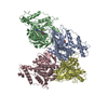
| ||||||||
|---|---|---|---|---|---|---|---|---|---|
| 1 |
| ||||||||
| 2 | 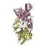
| ||||||||
| Unit cell |
| ||||||||
| Components on special symmetry positions |
|
-Components
| #1: Protein | Mass: 41167.598 Da / Num. of mol.: 4 / Mutation: E251A Source method: isolated from a genetically manipulated source Source: (gene. exp.) Acinetobacter baylyi ADP1 (bacteria) / Strain: ATCC 33305 / BD413 / ADP1 / Gene: ACIAD1282 / Production host: #2: Chemical | ChemComp-7ID / (   Type: L-peptide linking / Mass: 289.288 Da / Num. of mol.: 4 / Source method: obtained synthetically / Formula: C10H19N5O5 / Feature type: SUBJECT OF INVESTIGATION Type: L-peptide linking / Mass: 289.288 Da / Num. of mol.: 4 / Source method: obtained synthetically / Formula: C10H19N5O5 / Feature type: SUBJECT OF INVESTIGATION#3: Chemical | ChemComp-ZN /   Mass: 65.409 Da / Num. of mol.: 4 / Source method: obtained synthetically / Formula: Zn Mass: 65.409 Da / Num. of mol.: 4 / Source method: obtained synthetically / Formula: Zn#4: Chemical | ChemComp-MN /   Mass: 54.938 Da / Num. of mol.: 4 / Source method: isolated from a natural source / Formula: Mn Mass: 54.938 Da / Num. of mol.: 4 / Source method: isolated from a natural source / Formula: Mn#5: Water | ChemComp-HOH / |  Mass: 18.015 Da / Num. of mol.: 437 / Source method: isolated from a natural source / Formula: H2O Mass: 18.015 Da / Num. of mol.: 437 / Source method: isolated from a natural source / Formula: H2OHas ligand of interest | Y | |
|---|
-Experimental details
-Experiment
| Experiment | Method: X-RAY DIFFRACTION / Number of used crystals: 1 |
|---|
- Sample preparation
| Crystal | Density Matthews: 2.32 Å3/Da / Density % sol: 46.98 % |
|---|---|
| Crystal grow | Temperature: 277 K / Method: vapor diffusion, sitting drop / pH: 7.5 Details: 0.1 M bis-tris propane pH 7.5, 24% PEG3350, 0.2 M NaBr, 10 mM spermine and 10 mM LiCl |
-Data collection
| Diffraction | Mean temperature: 100 K / Serial crystal experiment: N |
|---|---|
| Diffraction source | Source: SYNCHROTRON / Site: ALS  / Beamline: 5.0.1 / Wavelength: 0.9774 Å / Beamline: 5.0.1 / Wavelength: 0.9774 Å |
| Detector | Type: DECTRIS PILATUS3 2M / Detector: PIXEL / Date: Jun 24, 2021 |
| Radiation | Protocol: SINGLE WAVELENGTH / Monochromatic (M) / Laue (L): M / Scattering type: x-ray |
| Radiation wavelength | Wavelength: 0.9774 Å / Relative weight: 1 |
| Reflection | Resolution: 2.24→85.6 Å / Num. obs: 73553 / % possible obs: 99.3 % / Redundancy: 2.8 % / CC1/2: 0.999 / Rmerge(I) obs: 0.07169 / Net I/σ(I): 6.68 |
| Reflection shell | Resolution: 2.4→2.49 Å / Rmerge(I) obs: 0.201 / Mean I/σ(I) obs: 0.3 / Num. unique obs: 6064 / CC1/2: 0.947 |
- Processing
| Software |
| |||||||||||||||||||||||||||||||||||||||||||||||||||||||||||||||||||||||||||||||||||||||||||||||||||||||||||||||||||||||||||||||||||||||||||||||||||||||||||||||||||||||||||||||||||||||||||||
|---|---|---|---|---|---|---|---|---|---|---|---|---|---|---|---|---|---|---|---|---|---|---|---|---|---|---|---|---|---|---|---|---|---|---|---|---|---|---|---|---|---|---|---|---|---|---|---|---|---|---|---|---|---|---|---|---|---|---|---|---|---|---|---|---|---|---|---|---|---|---|---|---|---|---|---|---|---|---|---|---|---|---|---|---|---|---|---|---|---|---|---|---|---|---|---|---|---|---|---|---|---|---|---|---|---|---|---|---|---|---|---|---|---|---|---|---|---|---|---|---|---|---|---|---|---|---|---|---|---|---|---|---|---|---|---|---|---|---|---|---|---|---|---|---|---|---|---|---|---|---|---|---|---|---|---|---|---|---|---|---|---|---|---|---|---|---|---|---|---|---|---|---|---|---|---|---|---|---|---|---|---|---|---|---|---|---|---|---|---|---|
| Refinement | Method to determine structure: MOLECULAR REPLACEMENT Starting model: Model generated using Rosetta Resolution: 2.24→49.04 Å / SU ML: 0.33 / Cross valid method: THROUGHOUT / σ(F): 1.38 / Phase error: 29.95 / Stereochemistry target values: ML
| |||||||||||||||||||||||||||||||||||||||||||||||||||||||||||||||||||||||||||||||||||||||||||||||||||||||||||||||||||||||||||||||||||||||||||||||||||||||||||||||||||||||||||||||||||||||||||||
| Solvent computation | Shrinkage radii: 0.9 Å / VDW probe radii: 1.1 Å / Solvent model: FLAT BULK SOLVENT MODEL | |||||||||||||||||||||||||||||||||||||||||||||||||||||||||||||||||||||||||||||||||||||||||||||||||||||||||||||||||||||||||||||||||||||||||||||||||||||||||||||||||||||||||||||||||||||||||||||
| Refinement step | Cycle: LAST / Resolution: 2.24→49.04 Å
| |||||||||||||||||||||||||||||||||||||||||||||||||||||||||||||||||||||||||||||||||||||||||||||||||||||||||||||||||||||||||||||||||||||||||||||||||||||||||||||||||||||||||||||||||||||||||||||
| Refine LS restraints |
| |||||||||||||||||||||||||||||||||||||||||||||||||||||||||||||||||||||||||||||||||||||||||||||||||||||||||||||||||||||||||||||||||||||||||||||||||||||||||||||||||||||||||||||||||||||||||||||
| LS refinement shell |
|
