 Movie
Movie Controller

[English] 日本語
 Yorodumi
- PDB-8eh2: Short-chain dehydrogenase/reductase (SDR) from Acinetobacter baumannii -
+ Open data

- Basic information
| Entry | Database: PDB / ID: 8eh2 | ||||||
|---|---|---|---|---|---|---|---|
| Title | Short-chain dehydrogenase/reductase (SDR) from Acinetobacter baumannii | ||||||
 Components Components | SDR family oxidoreductase | ||||||
 Keywords Keywords | OXIDOREDUCTASE / Short-chain / SDR | ||||||
| Function / homology |  Function and homology information Function and homology information3-oxoacyl-[acyl-carrier-protein] reductase (NADPH) activity / 3-oxoacyl-[acyl-carrier-protein] reductase Similarity search - Function | ||||||
| Biological species |  Acinetobacter baumannii (bacteria) Acinetobacter baumannii (bacteria) | ||||||
| Method |  X-RAY DIFFRACTION / SYNCHROTRON / MOLECULAR REPLACEMENT / Resolution: 2.4 Å X-RAY DIFFRACTION / SYNCHROTRON / MOLECULAR REPLACEMENT / Resolution: 2.4 Å | ||||||
 Authors Authors | Shaw, K. | ||||||
| Funding support | 1items
| ||||||
 Citation Citation | Journal: To Be Published Title: Short-chain dehydrogenase/reductase (SDR) from Acinetobacter baumannii Authors: Forwood, J.K. | ||||||
| History |
|
- Structure visualization
| Structure viewer | Molecule: MolmilJmol/JSmol |
|---|
- Downloads & links
-Download
| PDBx/mmCIF format | 8eh2.cif.gz | 231.9 KB | Display | PDBx/mmCIF format |
|---|---|---|---|---|
| PDB format | pdb8eh2.ent.gz | 153.1 KB | Display | PDB format |
| PDBx/mmJSON format | 8eh2.json.gz | Tree view | PDBx/mmJSON format | |
| Others |  Other downloads Other downloads |
-Validation report
| Arichive directory | https://data.pdbj.org/pub/pdb/validation_reports/eh/8eh2ftp://data.pdbj.org/pub/pdb/validation_reports/eh/8eh2 | HTTPS FTP |
|---|
-Related structure data
| Related structure data | 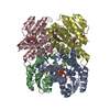 2d1yS S: Starting model for refinement |
|---|---|
| Similar structure data |
-Links
 PDBj
PDBj


- Assembly
| Deposited unit | 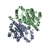
| |||||||||||||||||||||
|---|---|---|---|---|---|---|---|---|---|---|---|---|---|---|---|---|---|---|---|---|---|---|
| 1 | 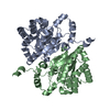
| |||||||||||||||||||||
| Unit cell |
| |||||||||||||||||||||
| Components on special symmetry positions |
| |||||||||||||||||||||
| Noncrystallographic symmetry (NCS) | NCS domain:
NCS domain segments: Ens-ID: 1 / Beg auth comp-ID: MET / Beg label comp-ID: MET / End auth comp-ID: LEU / End label comp-ID: LEU / Auth seq-ID: 1 - 259 / Label seq-ID: 1 - 259
|
-Components
| #1: Protein | Mass: 27827.480 Da / Num. of mol.: 2 Source method: isolated from a genetically manipulated source Source: (gene. exp.) Acinetobacter baumannii (bacteria) / Gene: F4T85_19375, FJU42_12090, HBK86_07635 / Production host: #2: Water | ChemComp-HOH / |  Mass: 18.015 Da / Num. of mol.: 201 / Source method: isolated from a natural source / Formula: H2O Mass: 18.015 Da / Num. of mol.: 201 / Source method: isolated from a natural source / Formula: H2O |
|---|
-Experimental details
-Experiment
| Experiment | Method: X-RAY DIFFRACTION / Number of used crystals: 1 |
|---|
- Sample preparation
| Crystal | Density Matthews: 2.27 Å3/Da / Density % sol: 45.81 % |
|---|---|
| Crystal grow | Temperature: 291 K / Method: vapor diffusion, hanging drop Details: 0.2M lithium chloride, 0.1M Tris pH 8.0, 20% (v/v) polyethylene glycol 8,000 |
-Data collection
| Diffraction | Mean temperature: 298 K / Ambient temp details: 80 / Serial crystal experiment: N |
|---|---|
| Diffraction source | Source: SYNCHROTRON / Site: Australian Synchrotron  / Beamline: MX2 / Wavelength: 0.9537 Å / Beamline: MX2 / Wavelength: 0.9537 Å |
| Detector | Type: DECTRIS EIGER X 16M / Detector: PIXEL / Date: Feb 24, 2018 |
| Radiation | Protocol: SINGLE WAVELENGTH / Monochromatic (M) / Laue (L): M / Scattering type: x-ray |
| Radiation wavelength | Wavelength: 0.9537 Å / Relative weight: 1 |
| Reflection | Resolution: 2.4→29.4 Å / Num. obs: 20194 / % possible obs: 99.9 % / Redundancy: 4.7 % / Biso Wilson estimate: 24.95 Å2 / CC1/2: 0.986 / Net I/σ(I): 6.9 |
| Reflection shell | Resolution: 2.4→2.49 Å / Num. unique obs: 2085 / CC1/2: 0.819 |
- Processing
| Software |
| |||||||||||||||||||||||||||||||||||||||||||||||||||||||||||||||
|---|---|---|---|---|---|---|---|---|---|---|---|---|---|---|---|---|---|---|---|---|---|---|---|---|---|---|---|---|---|---|---|---|---|---|---|---|---|---|---|---|---|---|---|---|---|---|---|---|---|---|---|---|---|---|---|---|---|---|---|---|---|---|---|---|
| Refinement | Method to determine structure: MOLECULAR REPLACEMENT Starting model: 2D1Y Resolution: 2.4→29.4 Å / SU ML: 0.2284 / Cross valid method: FREE R-VALUE / σ(F): 1.34 / Phase error: 22.5445 / Stereochemistry target values: CDL v1.2
| |||||||||||||||||||||||||||||||||||||||||||||||||||||||||||||||
| Solvent computation | Shrinkage radii: 0.9 Å / VDW probe radii: 1.11 Å / Solvent model: FLAT BULK SOLVENT MODEL | |||||||||||||||||||||||||||||||||||||||||||||||||||||||||||||||
| Displacement parameters | Biso mean: 31.76 Å2 | |||||||||||||||||||||||||||||||||||||||||||||||||||||||||||||||
| Refinement step | Cycle: LAST / Resolution: 2.4→29.4 Å
| |||||||||||||||||||||||||||||||||||||||||||||||||||||||||||||||
| Refine LS restraints |
| |||||||||||||||||||||||||||||||||||||||||||||||||||||||||||||||
| LS refinement shell |
|
