 Movie
Movie Controller


+ Open data

- Basic information
| Entry | Database: PDB / ID: 8egw | ||||||
|---|---|---|---|---|---|---|---|
| Title | Complex of Fat4(EC1-4) bound to Dchs1(EC1-3) | ||||||
 Components Components |
| ||||||
 Keywords Keywords | CELL ADHESION / Fat4 / Dachsous1 / Cadherin / Protocadherin / Adhesion / Fat / Dachsous | ||||||
| Function / homology |  Function and homology information Function and homology informationcondensed mesenchymal cell proliferation / septin cytoskeleton organization / mitral valve formation / cell migration involved in endocardial cushion formation / : / calcium-dependent cell-cell adhesion / hippo signaling / pattern specification process / post-anal tail morphogenesis / catenin complex ...condensed mesenchymal cell proliferation / septin cytoskeleton organization / mitral valve formation / cell migration involved in endocardial cushion formation / : / calcium-dependent cell-cell adhesion / hippo signaling / pattern specification process / post-anal tail morphogenesis / catenin complex / digestive tract development / heterophilic cell-cell adhesion / neural tube development / ossification involved in bone maturation / branching involved in ureteric bud morphogenesis / homophilic cell-cell adhesion / cochlea development / neurogenesis / protein localization to plasma membrane / cell-cell adhesion / beta-catenin binding / cerebral cortex development / apical part of cell / cell migration / gene expression / cadherin binding / calcium ion binding / extracellular exosome / membrane / plasma membrane Similarity search - Function | ||||||
| Biological species |  Homo sapiens (human) Homo sapiens (human) | ||||||
| Method |  X-RAY DIFFRACTION / SYNCHROTRON / MOLECULAR REPLACEMENT / Resolution: 2.3 Å X-RAY DIFFRACTION / SYNCHROTRON / MOLECULAR REPLACEMENT / Resolution: 2.3 Å | ||||||
 Authors Authors | Medina, E. / Luca, V.C. | ||||||
| Funding support |  United States, 1items United States, 1items
| ||||||
 Citation Citation | Journal: Nat Commun / Year: 2023 Title: Structure of the planar cell polarity cadherins Fat4 and Dachsous1. Authors: Medina, E. / Easa, Y. / Lester, D.K. / Lau, E.K. / Sprinzak, D. / Luca, V.C. | ||||||
| History |
|
- Structure visualization
| Structure viewer | Molecule: MolmilJmol/JSmol |
|---|
- Downloads & links
-Download
| PDBx/mmCIF format | 8egw.cif.gz | 317.7 KB | Display | PDBx/mmCIF format |
|---|---|---|---|---|
| PDB format | pdb8egw.ent.gz | 252.7 KB | Display | PDB format |
| PDBx/mmJSON format | 8egw.json.gz | Tree view | PDBx/mmJSON format | |
| Others |  Other downloads Other downloads |
-Validation report
| Arichive directory | https://data.pdbj.org/pub/pdb/validation_reports/eg/8egwftp://data.pdbj.org/pub/pdb/validation_reports/eg/8egw | HTTPS FTP |
|---|
-Related structure data
| Related structure data |  8egxC 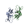 2a4cS 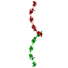 3q2wS  4zi8S  4zplS 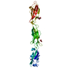 4zpoS  5iu9S S: Starting model for refinement C: citing same article ( |
|---|---|
| Similar structure data |
-Links
 PDBj
PDBj


- Assembly
| Deposited unit | 
| ||||||||
|---|---|---|---|---|---|---|---|---|---|
| 1 |
| ||||||||
| Unit cell |
|
-Components
-Protein , 2 types, 2 molecules AB
| #1: Protein | Mass: 33863.098 Da / Num. of mol.: 1 Source method: isolated from a genetically manipulated source Source: (gene. exp.) Homo sapiens (human) / Gene: DCHS1, CDH19, CDH25, FIB1, KIAA1773, PCDH16 / Production host:  Trichoplusia ni (cabbage looper) / References: UniProt: Q96JQ0 Trichoplusia ni (cabbage looper) / References: UniProt: Q96JQ0 |
|---|---|
| #2: Protein | Mass: 50777.395 Da / Num. of mol.: 1 Source method: isolated from a genetically manipulated source Source: (gene. exp.) Homo sapiens (human) / Gene: FAT4, CDHF14, FATJ, Nbla00548 / Production host: Trichoplusia ni (cabbage looper) / References: UniProt: Q6V0I7 |
-Sugars , 4 types, 5 molecules 
| #3: Polysaccharide | Source method: isolated from a genetically manipulated source #4: Polysaccharide | alpha-D-mannopyranose-(1-3)-beta-D-mannopyranose-(1-4)-2-acetamido-2-deoxy-beta-D-glucopyranose-(1- ...alpha-D-mannopyranose-(1-3)-beta-D-mannopyranose-(1-4)-2-acetamido-2-deoxy-beta-D-glucopyranose-(1-4)-[alpha-L-fucopyranose-(1-3)][alpha-L-fucopyranose-(1-6)]2-acetamido-2-deoxy-beta-D-glucopyranose | Source method: isolated from a genetically manipulated source #5: Polysaccharide | 2-acetamido-2-deoxy-beta-D-glucopyranose-(1-4)-[alpha-L-fucopyranose-(1-6)]2-acetamido-2-deoxy-beta- ...2-acetamido-2-deoxy-beta-D-glucopyranose-(1-4)-[alpha-L-fucopyranose-(1-6)]2-acetamido-2-deoxy-beta-D-glucopyranose | Source method: isolated from a genetically manipulated source #6: Sugar | ChemComp-NAG / |  Type: D-saccharide, beta linking / Mass: 221.208 Da / Num. of mol.: 1 / Source method: obtained synthetically / Formula: C8H15NO6 Type: D-saccharide, beta linking / Mass: 221.208 Da / Num. of mol.: 1 / Source method: obtained synthetically / Formula: C8H15NO6 |
|---|
-Non-polymers , 4 types, 314 molecules 


| #7: Chemical | ChemComp-CA /  Mass: 40.078 Da / Num. of mol.: 19 / Source method: obtained synthetically / Formula: Ca Mass: 40.078 Da / Num. of mol.: 19 / Source method: obtained synthetically / Formula: Ca#8: Chemical |  Mass: 136.989 Da / Num. of mol.: 3 / Source method: obtained synthetically / Formula: C2H6AsO2 Mass: 136.989 Da / Num. of mol.: 3 / Source method: obtained synthetically / Formula: C2H6AsO2#9: Chemical | ChemComp-EDO /  Mass: 62.068 Da / Num. of mol.: 12 / Source method: obtained synthetically / Formula: C2H6O2 Mass: 62.068 Da / Num. of mol.: 12 / Source method: obtained synthetically / Formula: C2H6O2#10: Water | ChemComp-HOH / | Mass: 18.015 Da / Num. of mol.: 280 / Source method: isolated from a natural source / Formula: H2O |
|---|
-Details
| Has ligand of interest | N |
|---|---|
| Has protein modification | Y |
-Experimental details
-Experiment
| Experiment | Method: X-RAY DIFFRACTION / Number of used crystals: 1 |
|---|
- Sample preparation
| Crystal | Density Matthews: 2.99 Å3/Da / Density % sol: 58.91 % |
|---|---|
| Crystal grow | Temperature: 293.15 K / Method: vapor diffusion, sitting drop Details: 0.08 M Magnesium Chloride; 0.1 M Sodium Cacodylate, pH 6.1; 20% Polyethylene Glycol 1000 |
-Data collection
| Diffraction | Mean temperature: 100 K / Serial crystal experiment: N |
|---|---|
| Diffraction source | Source: SYNCHROTRON / Site: APS / Beamline: 22-BM / Wavelength: 1 Å |
| Detector | Type: RAYONIX MX-300 / Detector: CCD / Date: Apr 11, 2019 |
| Radiation | Protocol: SINGLE WAVELENGTH / Monochromatic (M) / Laue (L): M / Scattering type: x-ray |
| Radiation wavelength | Wavelength: 1 Å / Relative weight: 1 |
| Reflection | Resolution: 2.204→49.085 Å / Num. obs: 50558 / % possible obs: 99.27 % / Redundancy: 3.4 % / CC1/2: 0.995 / Net I/σ(I): 8.87 |
| Reflection shell | Resolution: 2.204→2.283 Å / Num. unique obs: 5002 / CC1/2: 0.8 / % possible all: 98.24 |
- Processing
| Software |
| ||||||||||||||||||||||||||||||||||||||||||||||||||||||||||||||||||||||||||||||||||||||||||||||||||||||||||||||||||||||||||||||||||||||||||||||||||||||||||||||||||||||||||||||||||||||||||||||||||||||||
|---|---|---|---|---|---|---|---|---|---|---|---|---|---|---|---|---|---|---|---|---|---|---|---|---|---|---|---|---|---|---|---|---|---|---|---|---|---|---|---|---|---|---|---|---|---|---|---|---|---|---|---|---|---|---|---|---|---|---|---|---|---|---|---|---|---|---|---|---|---|---|---|---|---|---|---|---|---|---|---|---|---|---|---|---|---|---|---|---|---|---|---|---|---|---|---|---|---|---|---|---|---|---|---|---|---|---|---|---|---|---|---|---|---|---|---|---|---|---|---|---|---|---|---|---|---|---|---|---|---|---|---|---|---|---|---|---|---|---|---|---|---|---|---|---|---|---|---|---|---|---|---|---|---|---|---|---|---|---|---|---|---|---|---|---|---|---|---|---|---|---|---|---|---|---|---|---|---|---|---|---|---|---|---|---|---|---|---|---|---|---|---|---|---|---|---|---|---|---|---|---|---|
| Refinement | Method to determine structure: MOLECULAR REPLACEMENT Starting model: 2A4C, 4ZPL, 4ZI8, 4ZPO, 5IU9, 3Q2W Resolution: 2.3→49.083 Å / SU ML: 0.34 / Cross valid method: THROUGHOUT / σ(F): 1.35 / Phase error: 27.43 / Stereochemistry target values: ML
| ||||||||||||||||||||||||||||||||||||||||||||||||||||||||||||||||||||||||||||||||||||||||||||||||||||||||||||||||||||||||||||||||||||||||||||||||||||||||||||||||||||||||||||||||||||||||||||||||||||||||
| Solvent computation | Shrinkage radii: 0.9 Å / VDW probe radii: 1.11 Å / Solvent model: FLAT BULK SOLVENT MODEL | ||||||||||||||||||||||||||||||||||||||||||||||||||||||||||||||||||||||||||||||||||||||||||||||||||||||||||||||||||||||||||||||||||||||||||||||||||||||||||||||||||||||||||||||||||||||||||||||||||||||||
| Refinement step | Cycle: LAST / Resolution: 2.3→49.083 Å
| ||||||||||||||||||||||||||||||||||||||||||||||||||||||||||||||||||||||||||||||||||||||||||||||||||||||||||||||||||||||||||||||||||||||||||||||||||||||||||||||||||||||||||||||||||||||||||||||||||||||||
| Refine LS restraints |
| ||||||||||||||||||||||||||||||||||||||||||||||||||||||||||||||||||||||||||||||||||||||||||||||||||||||||||||||||||||||||||||||||||||||||||||||||||||||||||||||||||||||||||||||||||||||||||||||||||||||||
| LS refinement shell |
| ||||||||||||||||||||||||||||||||||||||||||||||||||||||||||||||||||||||||||||||||||||||||||||||||||||||||||||||||||||||||||||||||||||||||||||||||||||||||||||||||||||||||||||||||||||||||||||||||||||||||
| Refinement TLS params. | Method: refined / Refine-ID: X-RAY DIFFRACTION
| ||||||||||||||||||||||||||||||||||||||||||||||||||||||||||||||||||||||||||||||||||||||||||||||||||||||||||||||||||||||||||||||||||||||||||||||||||||||||||||||||||||||||||||||||||||||||||||||||||||||||
| Refinement TLS group |
|
