+ Open data
- Basic information
| Entry | Database: PDB / ID: 8eea | ||||||
|---|---|---|---|---|---|---|---|
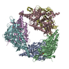
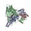
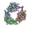
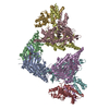
| Title | Structure of E.coli Septu (PtuAB) complex | ||||||
 Components Components |
| ||||||
 Keywords Keywords | IMMUNE SYSTEM / PtuA / PtuB Septu | ||||||
| Function / homology | Retron Ec78 putative HNH endonuclease-like / ADENOSINE-5'-TRIPHOSPHATE / TIGR02646 family protein Function and homology information Function and homology information | ||||||
| Biological species |  | ||||||
| Method | ELECTRON MICROSCOPY / single particle reconstruction / cryo EM / Resolution: 2.6 Å | ||||||
 Authors Authors | Shen, Z.F. / Fu, T.M. | ||||||
| Funding support | 1items
| ||||||
 Citation Citation |  Journal: Nat Struct Mol Biol / Year: 2024 Journal: Nat Struct Mol Biol / Year: 2024Title: PtuA and PtuB assemble into an inflammasome-like oligomer for anti-phage defense. Authors: Yuanyuan Li / Zhangfei Shen / Mengyuan Zhang / Xiao-Yuan Yang / Sean P Cleary / Jiale Xie / Ila A Marathe / Marius Kostelic / Jacelyn Greenwald / Anthony D Rish / Vicki H Wysocki / Chong ...Authors: Yuanyuan Li / Zhangfei Shen / Mengyuan Zhang / Xiao-Yuan Yang / Sean P Cleary / Jiale Xie / Ila A Marathe / Marius Kostelic / Jacelyn Greenwald / Anthony D Rish / Vicki H Wysocki / Chong Chen / Qiang Chen / Tian-Min Fu / Yamei Yu /   Abstract: Escherichia coli Septu system, an anti-phage defense system, comprises two components: PtuA and PtuB. PtuA contains an ATPase domain, while PtuB is predicted to function as a nuclease. Here we show ...Escherichia coli Septu system, an anti-phage defense system, comprises two components: PtuA and PtuB. PtuA contains an ATPase domain, while PtuB is predicted to function as a nuclease. Here we show that PtuA and PtuB form a stable complex with a 6:2 stoichiometry. Cryo-electron microscopy structure of PtuAB reveals a distinctive horseshoe-like configuration. PtuA adopts a hexameric arrangement, organized as an asymmetric trimer of dimers, contrasting the ring-like structure by other ATPases. Notably, the three pairs of PtuA dimers assume distinct conformations and fulfill unique roles in recruiting PtuB. Our functional assays have further illuminated the importance of the oligomeric assembly of PtuAB in anti-phage defense. Moreover, we have uncovered that ATP molecules can directly bind to PtuA and inhibit the activities of PtuAB. Together, the assembly and function of the Septu system shed light on understanding other ATPase-containing systems in bacterial immunity. | ||||||
| History |
|
- Structure visualization
| Structure viewer | Molecule:  Molmil Molmil Jmol/JSmol Jmol/JSmol |
|---|
- Downloads & links
- Download
| PDBx/mmCIF format |  8eea.cif.gz 8eea.cif.gz | 590 KB | Display |  PDBx/mmCIF format PDBx/mmCIF format |
|---|---|---|---|---|
| PDB format |  pdb8eea.ent.gz pdb8eea.ent.gz | 487.4 KB | Display |  PDB format PDB format |
| PDBx/mmJSON format |  8eea.json.gz 8eea.json.gz | Tree view |  PDBx/mmJSON format PDBx/mmJSON format | |
| Others |  Other downloads Other downloads |
-Validation report
| Arichive directory |  https://data.pdbj.org/pub/pdb/validation_reports/ee/8eea https://data.pdbj.org/pub/pdb/validation_reports/ee/8eea ftp://data.pdbj.org/pub/pdb/validation_reports/ee/8eea ftp://data.pdbj.org/pub/pdb/validation_reports/ee/8eea | HTTPS FTP |
|---|
-Related structure data
| Related structure data |  28049MC  8ee4C  8ee7C  8suxC M: map data used to model this data C: citing same article ( |
|---|---|
| Similar structure data | Similarity search - Function & homology  F&H Search F&H Search |
- Links
Links
- Assembly
| Deposited unit | 
|
|---|---|
| 1 |
|
- Components
| #1: Protein | Mass: 53189.656 Da / Num. of mol.: 6 Source method: isolated from a genetically manipulated source Source: (gene. exp.)   #2: Protein | Mass: 28136.291 Da / Num. of mol.: 2 Source method: isolated from a genetically manipulated source Source: (gene. exp.)   #3: Chemical | ChemComp-ATP / Has ligand of interest | Y | |
|---|
-Experimental details
-Experiment
| Experiment | Method: ELECTRON MICROSCOPY |
|---|---|
| EM experiment | Aggregation state: PARTICLE / 3D reconstruction method: single particle reconstruction |
- Sample preparation
| Component | Name: 6 PtuA and 2 PtuB form as a complex / Type: COMPLEX / Entity ID: #1-#2 / Source: RECOMBINANT |
|---|---|
| Source (natural) | Organism:  |
| Source (recombinant) | Organism:  |
| Buffer solution | pH: 7.5 |
| Specimen | Embedding applied: NO / Shadowing applied: NO / Staining applied: NO / Vitrification applied: YES |
| Vitrification | Cryogen name: ETHANE |
- Electron microscopy imaging
| Experimental equipment |  Model: Titan Krios / Image courtesy: FEI Company |
|---|---|
| Microscopy | Model: TFS KRIOS |
| Electron gun | Electron source:  FIELD EMISSION GUN / Accelerating voltage: 300 kV / Illumination mode: FLOOD BEAM FIELD EMISSION GUN / Accelerating voltage: 300 kV / Illumination mode: FLOOD BEAM |
| Electron lens | Mode: BRIGHT FIELD / Nominal defocus max: 2000 nm / Nominal defocus min: 500 nm / Cs: 2.7 mm |
| Image recording | Electron dose: 50 e/Å2 / Film or detector model: GATAN K3 (6k x 4k) |
- Processing
| CTF correction | Type: PHASE FLIPPING AND AMPLITUDE CORRECTION |
|---|---|
| 3D reconstruction | Resolution: 2.6 Å / Resolution method: FSC 0.143 CUT-OFF / Num. of particles: 2307662 / Symmetry type: POINT |
 Movie
Movie Controller


 PDBj
PDBj

