 Movie
Movie Controller


+ Open data

- Basic information
| Entry | Database: PDB / ID: 8eds | |||||||||
|---|---|---|---|---|---|---|---|---|---|---|
| Title | Escherichia coli pyruvate kinase (PykF) P70Q | |||||||||
 Components Components | Pyruvate kinase | |||||||||
 Keywords Keywords | TRANSFERASE / Pyruvate kinase / Long term evolution experiment / Kinase | |||||||||
| Function / homology |  Function and homology information Function and homology informationpyruvate kinase / pyruvate kinase activity / potassium ion binding / response to stress / kinase activity / magnesium ion binding / ATP binding Similarity search - Function | |||||||||
| Biological species |  | |||||||||
| Method |  X-RAY DIFFRACTION / SYNCHROTRON / MOLECULAR REPLACEMENT / Resolution: 2.1 Å X-RAY DIFFRACTION / SYNCHROTRON / MOLECULAR REPLACEMENT / Resolution: 2.1 Å | |||||||||
 Authors Authors | Donovan, K.A. / Coombes, D. / Dobson, R.C.J. / Cooper, T.F. | |||||||||
| Funding support |  United States, 2items United States, 2items
| |||||||||
 Citation Citation | Journal: To Be Published Title: Beneficial mutations occurring in E. coli pyruvate kinase afford new allosteric mechanisms leading to faster resumption of growth Authors: Donovan, K.A. / Coombes, D. / Dobson, R.C.J. / Cooper, T.F. | |||||||||
| History |
|
- Structure visualization
| Structure viewer | Molecule: MolmilJmol/JSmol |
|---|
- Downloads & links
-Download
| PDBx/mmCIF format | 8eds.cif.gz | 337.5 KB | Display | PDBx/mmCIF format |
|---|---|---|---|---|
| PDB format | pdb8eds.ent.gz | 281.3 KB | Display | PDB format |
| PDBx/mmJSON format | 8eds.json.gz | Tree view | PDBx/mmJSON format | |
| Others |  Other downloads Other downloads |
-Validation report
| Arichive directory | https://data.pdbj.org/pub/pdb/validation_reports/ed/8edsftp://data.pdbj.org/pub/pdb/validation_reports/ed/8eds | HTTPS FTP |
|---|
-Related structure data
| Related structure data | 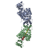 8edqC 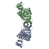 8edrC 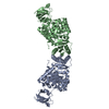 8edtC 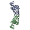 8eq0C  8eq1C  8eq3C  8eu4C  4yngS S: Starting model for refinement C: citing same article ( |
|---|---|
| Similar structure data |
-Links
 PDBj
PDBj
- Assembly
| Deposited unit | 
| ||||||||
|---|---|---|---|---|---|---|---|---|---|
| 1 | 
| ||||||||
| Unit cell |
|
-Components
| #1: Protein | Mass: 50796.312 Da / Num. of mol.: 2 / Mutation: P70T Source method: isolated from a genetically manipulated source Source: (gene. exp.) #2: Chemical |   Mass: 96.063 Da / Num. of mol.: 2 / Source method: obtained synthetically / Formula: SO4 Mass: 96.063 Da / Num. of mol.: 2 / Source method: obtained synthetically / Formula: SO4#3: Water | ChemComp-HOH / |  Mass: 18.015 Da / Num. of mol.: 187 / Source method: isolated from a natural source / Formula: H2O Mass: 18.015 Da / Num. of mol.: 187 / Source method: isolated from a natural source / Formula: H2OHas ligand of interest | N | |
|---|
-Experimental details
-Experiment
| Experiment | Method: X-RAY DIFFRACTION / Number of used crystals: 1 |
|---|
- Sample preparation
| Crystal | Density Matthews: 3.05 Å3/Da / Density % sol: 59.61 % |
|---|---|
| Crystal grow | Temperature: 293 K / Method: vapor diffusion, sitting drop Details: 0.1M (0.2M DL-Glutamic acid monohydrate; 0.2M DL-Alanine; 0.2M Glycine; 0.2M DL-Lysine monohydrochloride; 0.2M DL-Serine), 0.1M (1.0M Imidazole; MES monohydrate (acid)), 30% (40% v/v Glycerol; 20% w/v PEG 4000) |
-Data collection
| Diffraction | Mean temperature: 110 K / Serial crystal experiment: N |
|---|---|
| Diffraction source | Source: SYNCHROTRON / Site: Australian Synchrotron  / Beamline: MX1 / Wavelength: 0.9537 Å / Beamline: MX1 / Wavelength: 0.9537 Å |
| Detector | Type: ADSC QUANTUM 210r / Detector: CCD / Date: Sep 13, 2013 |
| Radiation | Protocol: SINGLE WAVELENGTH / Monochromatic (M) / Laue (L): M / Scattering type: x-ray |
| Radiation wavelength | Wavelength: 0.9537 Å / Relative weight: 1 |
| Reflection | Resolution: 2.1→44.8 Å / Num. obs: 72182 / % possible obs: 99.29 % / Redundancy: 1.9 % / CC1/2: 0.999 / Rmerge(I) obs: 0.039 / Net I/σ(I): 16.28 |
| Reflection shell | Resolution: 2.1→2.175 Å / Rmerge(I) obs: 0.3418 / Mean I/σ(I) obs: 2.7 / Num. unique obs: 6936 / CC1/2: 0.794 / % possible all: 96.64 |
- Processing
| Software |
| |||||||||||||||||||||||||||||||||||||||||||||||||||||||||||||||||||||||||||||||||||||||||||||||||||||||||||||||||||||||||||||||||||||
|---|---|---|---|---|---|---|---|---|---|---|---|---|---|---|---|---|---|---|---|---|---|---|---|---|---|---|---|---|---|---|---|---|---|---|---|---|---|---|---|---|---|---|---|---|---|---|---|---|---|---|---|---|---|---|---|---|---|---|---|---|---|---|---|---|---|---|---|---|---|---|---|---|---|---|---|---|---|---|---|---|---|---|---|---|---|---|---|---|---|---|---|---|---|---|---|---|---|---|---|---|---|---|---|---|---|---|---|---|---|---|---|---|---|---|---|---|---|---|---|---|---|---|---|---|---|---|---|---|---|---|---|---|---|---|
| Refinement | Method to determine structure: MOLECULAR REPLACEMENT Starting model: 4YNG Resolution: 2.1→44.8 Å / SU ML: 0.26 / Cross valid method: THROUGHOUT / σ(F): 1.9 / Phase error: 22.43 / Stereochemistry target values: MLHL
| |||||||||||||||||||||||||||||||||||||||||||||||||||||||||||||||||||||||||||||||||||||||||||||||||||||||||||||||||||||||||||||||||||||
| Solvent computation | Shrinkage radii: 0.9 Å / VDW probe radii: 1.11 Å / Solvent model: FLAT BULK SOLVENT MODEL | |||||||||||||||||||||||||||||||||||||||||||||||||||||||||||||||||||||||||||||||||||||||||||||||||||||||||||||||||||||||||||||||||||||
| Displacement parameters | Biso max: 114.12 Å2 / Biso mean: 38.5321 Å2 / Biso min: 18.56 Å2 | |||||||||||||||||||||||||||||||||||||||||||||||||||||||||||||||||||||||||||||||||||||||||||||||||||||||||||||||||||||||||||||||||||||
| Refinement step | Cycle: final / Resolution: 2.1→44.8 Å
| |||||||||||||||||||||||||||||||||||||||||||||||||||||||||||||||||||||||||||||||||||||||||||||||||||||||||||||||||||||||||||||||||||||
| LS refinement shell | Refine-ID: X-RAY DIFFRACTION / Rfactor Rfree error: 0 / Total num. of bins used: 18
|
