 Movie
Movie Controller

[English] 日本語
 Yorodumi
- PDB-8ec3: The crystal structure of the complement inhibitory domain of Borr... -
+ Open data

- Basic information
| Entry | Database: PDB / ID: 8ec3 | ||||||
|---|---|---|---|---|---|---|---|
| Title | The crystal structure of the complement inhibitory domain of Borrelia hermsii FbpC. | ||||||
 Components Components | Fibronectin-binding protein | ||||||
 Keywords Keywords | IMMUNE SYSTEM / Borrelia hermsii / complement / inhibitor | ||||||
| Function / homology | Prokaryotic membrane lipoprotein lipid attachment site profile. / metal ion binding / Fibronectin-binding protein Function and homology information Function and homology information | ||||||
| Biological species |  Borrelia hermsii HS1 (bacteria) Borrelia hermsii HS1 (bacteria) | ||||||
| Method |  X-RAY DIFFRACTION / SYNCHROTRON / MOLECULAR REPLACEMENT / Resolution: 1.5 Å X-RAY DIFFRACTION / SYNCHROTRON / MOLECULAR REPLACEMENT / Resolution: 1.5 Å | ||||||
 Authors Authors | Booth, C.E. / Garcia, B.L. | ||||||
| Funding support |  United States, 1items United States, 1items
| ||||||
 Citation Citation | Journal: J.Biol.Chem. / Year: 2023 Title: Conformational dynamics of complement protease C1r inhibitor proteins from Lyme disease- and relapsing fever-causing spirochetes. Authors: Roy, S. / Booth Jr., C.E. / Powell-Pierce, A.D. / Schulz, A.M. / Skare, J.T. / Garcia, B.L. #1: Journal: Biorxiv / Year: 2023 Title: "Conformational dynamics of C1r inhibitor proteins from Lyme disease and relapsing fever spirochetes". Authors: Roy, S. / Booth, C.E. / Powell-Pierce, A.D. / Schulz, A.M. / Skare, J.T. / Garcia, B.L. | ||||||
| History |
|
- Structure visualization
| Structure viewer | Molecule: MolmilJmol/JSmol |
|---|
- Downloads & links
-Download
| PDBx/mmCIF format | 8ec3.cif.gz | 80.4 KB | Display | PDBx/mmCIF format |
|---|---|---|---|---|
| PDB format | pdb8ec3.ent.gz | 55.9 KB | Display | PDB format |
| PDBx/mmJSON format | 8ec3.json.gz | Tree view | PDBx/mmJSON format | |
| Others |  Other downloads Other downloads |
-Validation report
| Arichive directory | https://data.pdbj.org/pub/pdb/validation_reports/ec/8ec3ftp://data.pdbj.org/pub/pdb/validation_reports/ec/8ec3 | HTTPS FTP |
|---|
-Related structure data
| Similar structure data |
|---|
-Links
 PDBj
PDBj


- Assembly
| Deposited unit | 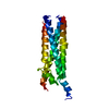
| ||||||||||||
|---|---|---|---|---|---|---|---|---|---|---|---|---|---|
| 1 |
| ||||||||||||
| Unit cell |
|
-Components
| #1: Protein | Mass: 18881.260 Da / Num. of mol.: 1 Source method: isolated from a genetically manipulated source Source: (gene. exp.) Borrelia hermsii HS1 (bacteria) / Gene: cihC, A0V01_04335, BHA007 / Plasmid: pT7HMT / Production host: |
|---|---|
| #2: Chemical | ChemComp-MG /   Mass: 24.305 Da / Num. of mol.: 1 / Source method: obtained synthetically / Formula: Mg Mass: 24.305 Da / Num. of mol.: 1 / Source method: obtained synthetically / Formula: Mg |
| #3: Water | ChemComp-HOH /  Mass: 18.015 Da / Num. of mol.: 157 / Source method: isolated from a natural source / Formula: H2O Mass: 18.015 Da / Num. of mol.: 157 / Source method: isolated from a natural source / Formula: H2O |
| Has ligand of interest | N |
-Experimental details
-Experiment
| Experiment | Method: X-RAY DIFFRACTION / Number of used crystals: 1 |
|---|
- Sample preparation
| Crystal | Density Matthews: 1.99 Å3/Da / Density % sol: 38.2 % / Description: Large birefringent plates |
|---|---|
| Crystal grow | Temperature: 298.15 K / Method: vapor diffusion, sitting drop / pH: 5.5 Details: Crystals were grown in the following condition containing 0.2M magnesium (II) chloride, 0.1M bis-tris (pH 5.5), and 25% PEG 3,350. Crystals were cryoprotected in a buffer containing 0.2M ...Details: Crystals were grown in the following condition containing 0.2M magnesium (II) chloride, 0.1M bis-tris (pH 5.5), and 25% PEG 3,350. Crystals were cryoprotected in a buffer containing 0.2M magnesium (II) chloride, 0.1M sodium citrate (pH 5.4), 25% PEG 3,350, and 10% glycerol. |
-Data collection
| Diffraction | Mean temperature: 93 K / Serial crystal experiment: N |
|---|---|
| Diffraction source | Source: SYNCHROTRON / Site: APS / Beamline: 22-BM / Wavelength: 1 Å |
| Detector | Type: MARMOSAIC 300 mm CCD / Detector: CCD / Date: Nov 29, 2021 |
| Radiation | Monochromator: Si(111) / Protocol: SINGLE WAVELENGTH / Monochromatic (M) / Laue (L): M / Scattering type: x-ray |
| Radiation wavelength | Wavelength: 1 Å / Relative weight: 1 |
| Reflection | Resolution: 1.5→31.28 Å / Num. obs: 23546 / % possible obs: 98.7 % / Redundancy: 7 % / Biso Wilson estimate: 21.7 Å2 / CC1/2: 0.989 / CC star: 0.997 / Rpim(I) all: 0.032 / Rrim(I) all: 0.087 / Net I/σ(I): 19.4 |
| Reflection shell | Resolution: 1.5→1.55 Å / Mean I/σ(I) obs: 2 / Num. unique obs: 2109 / CC1/2: 0.87 / CC star: 0.965 / Rpim(I) all: 0.218 / Rrim(I) all: 0.509 / % possible all: 89.2 |
- Processing
| Software |
| |||||||||||||||||||||||||||||||||||||||||||||||||||||||||||||||||||||||||||||||||||||||||||||||||||||||||
|---|---|---|---|---|---|---|---|---|---|---|---|---|---|---|---|---|---|---|---|---|---|---|---|---|---|---|---|---|---|---|---|---|---|---|---|---|---|---|---|---|---|---|---|---|---|---|---|---|---|---|---|---|---|---|---|---|---|---|---|---|---|---|---|---|---|---|---|---|---|---|---|---|---|---|---|---|---|---|---|---|---|---|---|---|---|---|---|---|---|---|---|---|---|---|---|---|---|---|---|---|---|---|---|---|---|---|
| Refinement | Method to determine structure: MOLECULAR REPLACEMENT Starting model: AlphaFold Resolution: 1.5→31.28 Å / SU ML: 0.1587 / Cross valid method: FREE R-VALUE / σ(F): 0 / Phase error: 22.9463 Stereochemistry target values: GeoStd + Monomer Library + CDL v1.2
| |||||||||||||||||||||||||||||||||||||||||||||||||||||||||||||||||||||||||||||||||||||||||||||||||||||||||
| Solvent computation | Shrinkage radii: 0.9 Å / VDW probe radii: 1.11 Å / Solvent model: FLAT BULK SOLVENT MODEL | |||||||||||||||||||||||||||||||||||||||||||||||||||||||||||||||||||||||||||||||||||||||||||||||||||||||||
| Displacement parameters | Biso mean: 31.39 Å2 | |||||||||||||||||||||||||||||||||||||||||||||||||||||||||||||||||||||||||||||||||||||||||||||||||||||||||
| Refinement step | Cycle: LAST / Resolution: 1.5→31.28 Å
| |||||||||||||||||||||||||||||||||||||||||||||||||||||||||||||||||||||||||||||||||||||||||||||||||||||||||
| Refine LS restraints |
| |||||||||||||||||||||||||||||||||||||||||||||||||||||||||||||||||||||||||||||||||||||||||||||||||||||||||
| LS refinement shell |
|
