 Movie
Movie Controller


+ Open data

- Basic information
| Entry | Database: PDB / ID: 8e7y | ||||||
|---|---|---|---|---|---|---|---|
| Title | RsTSPO A138F with two heme bound | ||||||
 Components Components | Tryptophan-rich sensory protein | ||||||
 Keywords Keywords | MEMBRANE PROTEIN / Translocator Protein 18 kD / TSPO | ||||||
| Function / homology |  Function and homology information Function and homology informationtetrapyrrole metabolic process / tetrapyrrole binding / lipid binding / identical protein binding / membrane / plasma membrane Similarity search - Function | ||||||
| Biological species |  Cereibacter sphaeroides (bacteria) Cereibacter sphaeroides (bacteria) | ||||||
| Method |  X-RAY DIFFRACTION / SYNCHROTRON / MOLECULAR REPLACEMENT / Resolution: 2.3 Å X-RAY DIFFRACTION / SYNCHROTRON / MOLECULAR REPLACEMENT / Resolution: 2.3 Å | ||||||
 Authors Authors | Liu, J. / Hiser, C. / Li, F. / Garavito, R. / Ferguson-Miller, S. | ||||||
| Funding support |  United States, 1items United States, 1items
| ||||||
 Citation Citation | Journal: Biochemistry / Year: 2023 Title: New TSPO Crystal Structures of Mutant and Heme-Bound Forms with Altered Flexibility, Ligand Binding, and Porphyrin Degradation Activity. Authors: Liu, J. / Hiser, C. / Li, F. / Hall, R. / Garavito, R.M. / Ferguson-Miller, S. | ||||||
| History |
|
- Structure visualization
| Structure viewer | Molecule: MolmilJmol/JSmol |
|---|
- Downloads & links
-Download
| PDBx/mmCIF format | 8e7y.cif.gz | 82 KB | Display | PDBx/mmCIF format |
|---|---|---|---|---|
| PDB format | pdb8e7y.ent.gz | 59.7 KB | Display | PDB format |
| PDBx/mmJSON format | 8e7y.json.gz | Tree view | PDBx/mmJSON format | |
| Others |  Other downloads Other downloads |
-Validation report
| Summary document | 8e7y_validation.pdf.gz | 1.8 MB | Display | wwPDB validaton report |
|---|---|---|---|---|
| Full document | 8e7y_full_validation.pdf.gz | 1.8 MB | Display | |
| Data in XML | 8e7y_validation.xml.gz | 15 KB | Display | |
| Data in CIF | 8e7y_validation.cif.gz | 18.5 KB | Display | |
| Arichive directory | https://data.pdbj.org/pub/pdb/validation_reports/e7/8e7yftp://data.pdbj.org/pub/pdb/validation_reports/e7/8e7y | HTTPS FTP |
-Related structure data
| Related structure data |  8e7wC  8e7xC  8e7zC  4uc1S S: Starting model for refinement C: citing same article ( |
|---|---|
| Similar structure data |
-Links
 PDBj
PDBj


- Assembly
| Deposited unit | 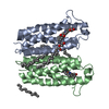
| |||||||||||||||||||||||||||||||||||||||||||||||||||||||||||||||||||||||||||||||
|---|---|---|---|---|---|---|---|---|---|---|---|---|---|---|---|---|---|---|---|---|---|---|---|---|---|---|---|---|---|---|---|---|---|---|---|---|---|---|---|---|---|---|---|---|---|---|---|---|---|---|---|---|---|---|---|---|---|---|---|---|---|---|---|---|---|---|---|---|---|---|---|---|---|---|---|---|---|---|---|---|
| 1 |
| |||||||||||||||||||||||||||||||||||||||||||||||||||||||||||||||||||||||||||||||
| Unit cell |
| |||||||||||||||||||||||||||||||||||||||||||||||||||||||||||||||||||||||||||||||
| Noncrystallographic symmetry (NCS) | NCS domain:
NCS domain segments:
|