National Institutes of Health/National Institute of General Medical Sciences (NIH/NIGMS)
GM136508
米国
引用
ジャーナル: J Struct Biol / 年: 2022 タイトル: Electron-counting MicroED data with the K2 and K3 direct electron detectors. 著者: Max T B Clabbers / Michael W Martynowycz / Johan Hattne / Brent L Nannenga / Tamir Gonen / 要旨: Microcrystal electron diffraction (MicroED) uses electron cryo-microscopy (cryo-EM) to collect diffraction data from small crystals during continuous rotation of the sample. As a result of advances ...Microcrystal electron diffraction (MicroED) uses electron cryo-microscopy (cryo-EM) to collect diffraction data from small crystals during continuous rotation of the sample. As a result of advances in hardware as well as methods development, the data quality has continuously improved over the past decade, to the point where even macromolecular structures can be determined ab initio. Detectors suitable for electron diffraction should ideally have fast readout to record data in movie mode, and high sensitivity at low exposure rates to accurately report the intensities. Direct electron detectors are commonly used in cryo-EM imaging for their sensitivity and speed, but despite their availability are generally not used in diffraction. Primary concerns with diffraction experiments are the dynamic range and coincidence loss, which will corrupt the measurement if the flux exceeds the count rate of the detector. Here, we describe instrument setup and low-exposure MicroED data collection in electron-counting mode using K2 and K3 direct electron detectors and show that the integrated intensities can be effectively used to solve structures of two macromolecules between 1.2 Å and 2.8 Å resolution. Even though a beam stop was not used with the K3 studies we did not observe damage to the camera. As these cameras are already available in many cryo-EM facilities, this provides opportunities for users who do not have access to dedicated facilities for MicroED.
名称: REFMAC / バージョン: 5.8.0267 / 分類: 精密化 / Contact author: Garib N. Murshudov / Contact author email: garib[at]mrc-lmb.cam.ac.uk / 日付: 2020-24-08 解説: (un)restrained refinement or idealisation of macromolecular structures
EMソフトウェア
ID
名称
バージョン
カテゴリ
11
AIMLESS
crystallographymerging
12
REFMAC
5.8.0267
3次元再構成
13
REFMAC
5.8.0267
モデル精密化
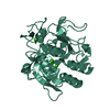
EM 3D crystal entity
∠α: 87.85 ° / ∠β: 108.85 ° / ∠γ: 112.6 ° / A: 26.38 Å / B: 30.76 Å / C: 33 Å / 空間群名: P-1 / 空間群番号: 2
CTF補正
タイプ: NONE
3次元再構成
解像度の算出法: DIFFRACTION PATTERN/LAYERLINES / 対称性のタイプ: 3D CRYSTAL
原子モデル構築
B value: 12.94 / プロトコル: AB INITIO MODEL / 空間: RECIPROCAL / Target criteria: Maximum likelihood
精密化
解像度: 1.2→31.076 Å / Cor.coef. Fo:Fc: 0.97 / Cor.coef. Fo:Fc free: 0.944 / SU B: 3.756 / SU ML: 0.069 / 交差検証法: THROUGHOUT / ESU R: 0.068 / ESU R Free: 0.068 / 詳細: Hydrogens have been added in their riding positions
Rfactor
反射数
%反射
Rfree
0.242
1186
4.838 %
Rwork
0.1813
23326
-
all
0.184
-
-
obs
-
24512
86.839 %
溶媒の処理
イオンプローブ半径: 0.8 Å / 減衰半径: 0.8 Å / VDWプローブ半径: 1.2 Å / 溶媒モデル: MASK BULK SOLVENT
 ムービー
ムービー コントローラー


 データを開く
 基本情報
基本情報 要素
要素 キーワード
キーワード 機能・相同性情報
 データ登録者
 米国, 2件
  引用
引用 構造の表示
構造の表示 ダウンロードとリンク
 その他のダウンロード

 PDBj
PDBj


 集合体
集合体

 分子量: 62.005 Da / 分子数: 1 / 由来タイプ: 合成 / 式: NO3
 分子量: 18.015 Da / 分子数: 118 / 由来タイプ: 天然 / 式: H2O
 試料調製
試料調製
 FIELD EMISSION GUN / 加速電圧: 300 kV / 照射モード: FLOOD BEAM
 解析
解析