National Institutes of Health/National Institute of General Medical Sciences (NIH/NIGMS)
GM136508
United States
Citation
Journal: J Struct Biol / Year: 2022 Title: Electron-counting MicroED data with the K2 and K3 direct electron detectors. Authors: Max T B Clabbers / Michael W Martynowycz / Johan Hattne / Brent L Nannenga / Tamir Gonen / Abstract: Microcrystal electron diffraction (MicroED) uses electron cryo-microscopy (cryo-EM) to collect diffraction data from small crystals during continuous rotation of the sample. As a result of advances ...Microcrystal electron diffraction (MicroED) uses electron cryo-microscopy (cryo-EM) to collect diffraction data from small crystals during continuous rotation of the sample. As a result of advances in hardware as well as methods development, the data quality has continuously improved over the past decade, to the point where even macromolecular structures can be determined ab initio. Detectors suitable for electron diffraction should ideally have fast readout to record data in movie mode, and high sensitivity at low exposure rates to accurately report the intensities. Direct electron detectors are commonly used in cryo-EM imaging for their sensitivity and speed, but despite their availability are generally not used in diffraction. Primary concerns with diffraction experiments are the dynamic range and coincidence loss, which will corrupt the measurement if the flux exceeds the count rate of the detector. Here, we describe instrument setup and low-exposure MicroED data collection in electron-counting mode using K2 and K3 direct electron detectors and show that the integrated intensities can be effectively used to solve structures of two macromolecules between 1.2 Å and 2.8 Å resolution. Even though a beam stop was not used with the K3 studies we did not observe damage to the camera. As these cameras are already available in many cryo-EM facilities, this provides opportunities for users who do not have access to dedicated facilities for MicroED.
Cryogen: NITROGEN / Specimen holder model: FEI TITAN KRIOS AUTOGRID HOLDER / Temperature (max): 90 K / Temperature (min): 77 K
Image recording
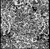
Average exposure time: 0.1 sec. / Electron dose: 0.001 e/Å2 / Film or detector model: GATAN K3 (6k x 4k) / Num. of diffraction images: 5600 / Num. of grids imaged: 1 / Num. of real images: 1
Image scans
Sampling size: 5 µm / Width: 5760 / Height: 4092
EM diffraction
Camera length: 373 mm
EM diffraction shell
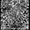
Resolution: 0.87→0.9 Å / Fourier space coverage: 37.64 % / Multiplicity: 2.1 / Num. of structure factors: 2783 / Phase residual: 30 °
EM diffraction stats
Fourier space coverage: 87.58 % / High resolution: 0.87 Å / Num. of intensities measured: 569407 / Num. of structure factors: 64974 / Phase error: 30 ° / Phase error rejection criteria: None / Rmerge: 0.236 / Rsym: 0.073
-
Processing
Software
Name: REFMAC / Version: 5.8.0267 / Classification: refinement / Contact author: Garib N. Murshudov / Contact author email: garib[at]mrc-lmb.cam.ac.uk / Date: 2020-24-08 Description: (un)restrained refinement or idealisation of macromolecular structures
EM software
ID
Name
Version
Category
11
AIMLESS
crystallographymerging
12
REFMAC
5.8.0267
3Dreconstruction
13
REFMAC
5.8.0267
modelrefinement
EM 3D crystal entity
∠α: 87.85 ° / ∠β: 108.85 ° / ∠γ: 112.6 ° / A: 26.38 Å / B: 30.76 Å / C: 33 Å / Space group name: P-1 / Space group num: 2
CTF correction
Type: NONE
3D reconstruction
Resolution method: DIFFRACTION PATTERN/LAYERLINES / Symmetry type: 3D CRYSTAL
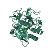
Atomic model building
B value: 12.94 / Protocol: AB INITIO MODEL / Space: RECIPROCAL / Target criteria: Maximum likelihood
Refinement
Resolution: 1.2→31.076 Å / Cor.coef. Fo:Fc: 0.97 / Cor.coef. Fo:Fc free: 0.944 / SU B: 3.756 / SU ML: 0.069 / Cross valid method: THROUGHOUT / ESU R: 0.068 / ESU R Free: 0.068 Details: Hydrogens have been added in their riding positions
Rfactor
Num. reflection
% reflection
Rfree
0.242
1186
4.838 %
Rwork
0.1813
23326
-
all
0.184
-
-
obs
-
24512
86.839 %
Solvent computation
Ion probe radii: 0.8 Å / Shrinkage radii: 0.8 Å / VDW probe radii: 1.2 Å / Solvent model: MASK BULK SOLVENT
 Movie
Movie Controller


 Open data
 Basic information
 Components
 Keywords
 Function and homology information
 Authors
 United States, 2items
  Citation
 Structure visualization
 Downloads & links
 Other downloads
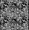

 PDBj
PDBj


 Assembly

 Mass: 62.005 Da / Num. of mol.: 1 / Source method: obtained synthetically / Formula: NO3
 Mass: 18.015 Da / Num. of mol.: 118 / Source method: isolated from a natural source / Formula: H2O
 Sample preparation
 FIELD EMISSION GUN / Accelerating voltage: 300 kV / Illumination mode: FLOOD BEAM
 Processing