 Movie
Movie Controller

[English] 日本語
 Yorodumi
- PDB-8e4t: Crystal structure of the kinase domain of RTKC8 from the choanofl... -
+ Open data

- Basic information
| Entry | Database: PDB / ID: 8e4t | ||||||
|---|---|---|---|---|---|---|---|
| Title | Crystal structure of the kinase domain of RTKC8 from the choanoflagellate Monosiga brevicollis | ||||||
 Components Components | RTKC8 Kinase domain | ||||||
 Keywords Keywords | TRANSFERASE / Receptor Tyrosine Kinase Staurosporine | ||||||
| Function / homology |  Function and homology information Function and homology informationtransmembrane receptor protein tyrosine kinase activity / receptor complex / ATP binding / plasma membrane Similarity search - Function | ||||||
| Biological species |  | ||||||
| Method |  X-RAY DIFFRACTION / SYNCHROTRON / MOLECULAR REPLACEMENT / Resolution: 1.95 Å X-RAY DIFFRACTION / SYNCHROTRON / MOLECULAR REPLACEMENT / Resolution: 1.95 Å | ||||||
 Authors Authors | Bajaj, T. / Gee, C.L. / Kuriyan, J. | ||||||
| Funding support |  United States, 1items United States, 1items
| ||||||
 Citation Citation | Journal: Plos One / Year: 2023 Title: Crystal structure of the kinase domain of a receptor tyrosine kinase from a choanoflagellate, Monosiga brevicollis. Authors: Bajaj, T. / Kuriyan, J. / Gee, C.L. | ||||||
| History |
|
- Structure visualization
| Structure viewer | Molecule: MolmilJmol/JSmol |
|---|
- Downloads & links
-Download
| PDBx/mmCIF format | 8e4t.cif.gz | 85.1 KB | Display | PDBx/mmCIF format |
|---|---|---|---|---|
| PDB format | pdb8e4t.ent.gz | 50.4 KB | Display | PDB format |
| PDBx/mmJSON format | 8e4t.json.gz | Tree view | PDBx/mmJSON format | |
| Others |  Other downloads Other downloads |
-Validation report
| Summary document | 8e4t_validation.pdf.gz | 1.2 MB | Display | wwPDB validaton report |
|---|---|---|---|---|
| Full document | 8e4t_full_validation.pdf.gz | 1.2 MB | Display | |
| Data in XML | 8e4t_validation.xml.gz | 13 KB | Display | |
| Data in CIF | 8e4t_validation.cif.gz | 17.8 KB | Display | |
| Arichive directory | https://data.pdbj.org/pub/pdb/validation_reports/e4/8e4tftp://data.pdbj.org/pub/pdb/validation_reports/e4/8e4t | HTTPS FTP |
-Related structure data
| Related structure data |  4ueuS S: Starting model for refinement |
|---|---|
| Similar structure data |
-Links
 PDBj
PDBj- Assembly
| Deposited unit | 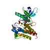
| ||||||||||
|---|---|---|---|---|---|---|---|---|---|---|---|
| 1 |
| ||||||||||
| Unit cell |
|
-Components
| #1: Protein | Mass: 31845.404 Da / Num. of mol.: 1 Source method: isolated from a genetically manipulated source Source: (gene. exp.)   Spodoptera frugiperda (fall armyworm) / References: UniProt: A9VBW0 Spodoptera frugiperda (fall armyworm) / References: UniProt: A9VBW0 | ||||||
|---|---|---|---|---|---|---|---|
| #2: Chemical |   Mass: 466.531 Da / Num. of mol.: 2 / Source method: obtained synthetically / Formula: C28H26N4O3 / Feature type: SUBJECT OF INVESTIGATION / Comment: antibiotic*YM Mass: 466.531 Da / Num. of mol.: 2 / Source method: obtained synthetically / Formula: C28H26N4O3 / Feature type: SUBJECT OF INVESTIGATION / Comment: antibiotic*YM#3: Chemical | ChemComp-PO4 / |   Mass: 94.971 Da / Num. of mol.: 1 / Source method: obtained synthetically / Formula: PO4 Mass: 94.971 Da / Num. of mol.: 1 / Source method: obtained synthetically / Formula: PO4#4: Water | ChemComp-HOH / |  Mass: 18.015 Da / Num. of mol.: 100 / Source method: isolated from a natural source / Formula: H2O Mass: 18.015 Da / Num. of mol.: 100 / Source method: isolated from a natural source / Formula: H2OHas ligand of interest | Y | |
-Experimental details
-Experiment
| Experiment | Method: X-RAY DIFFRACTION / Number of used crystals: 1 |
|---|
- Sample preparation
| Crystal | Density Matthews: 2.27 Å3/Da / Density % sol: 45.92 % Description: Flat triangular shaped, size 10x70x100x microns |
|---|---|
| Crystal grow | Temperature: 293 K / Method: vapor diffusion, sitting drop / pH: 6 Details: 0.1M SPG (Succinic acid, sodium dihydrogen phosphate and glycine) buffer pH 6.0, 25% (w/v) PEG 1500 Mixed 1:1 Protein: 10-15mg/mL in 50mM Tris pH 8.0, 200mM NaCl, 10% Glycerol, 0.5mM TCEP. |
-Data collection
| Diffraction | Mean temperature: 100 K / Serial crystal experiment: N |
|---|---|
| Diffraction source | Source: SYNCHROTRON / Site: ALS / Beamline: 8.2.1 / Wavelength: 0.9998 Å |
| Detector | Type: ADSC QUANTUM 315r / Detector: CCD / Date: Mar 5, 2019 |
| Radiation | Monochromator: Si111 / Protocol: SINGLE WAVELENGTH / Monochromatic (M) / Laue (L): M / Scattering type: x-ray |
| Radiation wavelength | Wavelength: 0.9998 Å / Relative weight: 1 |
| Reflection | Resolution: 1.95→48.555 Å / Num. obs: 20593 / % possible obs: 99.9 % / Redundancy: 26.2 % / Biso Wilson estimate: 32.71 Å2 / CC1/2: 0.998 / Rmerge(I) obs: 0.231 / Rpim(I) all: 0.046 / Rrim(I) all: 0.235 / Χ2: 0.95 / Net I/σ(I): 12.4 |
| Reflection shell | Resolution: 1.95→2 Å / Redundancy: 17 % / Rmerge(I) obs: 3.648 / Mean I/σ(I) obs: 0.9 / Num. unique obs: 1414 / CC1/2: 0.455 / Rpim(I) all: 0.883 / Rrim(I) all: 3.758 / Χ2: 0.76 / % possible all: 99.3 |
- Processing
| Software |
| ||||||||||||||||||||||||||||||||||||||||||||||||||||||||
|---|---|---|---|---|---|---|---|---|---|---|---|---|---|---|---|---|---|---|---|---|---|---|---|---|---|---|---|---|---|---|---|---|---|---|---|---|---|---|---|---|---|---|---|---|---|---|---|---|---|---|---|---|---|---|---|---|---|
| Refinement | Method to determine structure: MOLECULAR REPLACEMENT Starting model: 4UEU Resolution: 1.95→48.55 Å / SU ML: 0.2722 / Cross valid method: FREE R-VALUE / σ(F): 1.34 / Phase error: 25.9238 Stereochemistry target values: GeoStd + Monomer Library + CDL v1.2
| ||||||||||||||||||||||||||||||||||||||||||||||||||||||||
| Solvent computation | Shrinkage radii: 0.9 Å / VDW probe radii: 1.11 Å / Solvent model: FLAT BULK SOLVENT MODEL | ||||||||||||||||||||||||||||||||||||||||||||||||||||||||
| Displacement parameters | Biso mean: 35.07 Å2 | ||||||||||||||||||||||||||||||||||||||||||||||||||||||||
| Refinement step | Cycle: LAST / Resolution: 1.95→48.55 Å
| ||||||||||||||||||||||||||||||||||||||||||||||||||||||||
| Refine LS restraints |
| ||||||||||||||||||||||||||||||||||||||||||||||||||||||||
| LS refinement shell |
|
