host cell periplasmic space / virus tail, tube / viral portal complex / symbiont genome ejection through host cell envelope, short tail mechanism / viral DNA genome packaging / virus tail, fiber / adhesion receptor-mediated virion attachment to host cell / virion component / symbiont entry into host cell / virion attachment to host cell ...host cell periplasmic space / virus tail, tube / viral portal complex / symbiont genome ejection through host cell envelope, short tail mechanism / viral DNA genome packaging / virus tail, fiber / adhesion receptor-mediated virion attachment to host cell / virion component / symbiont entry into host cell / virion attachment to host cell / host cell plasma membrane / identical protein binding Similarity search - Function
Internal virion protein Gp15 / Phage T7 Internal virion protein gp15 / Internal virion protein Gp14 / T7 virus internal virion protein gp14 family / : / Tail fibre protein gp37 trimerization region / Bacteriophage T7, Gp17, C-terminal / Tail fibre protein gp37 C terminal domain / : / Bacteriophage nozzle protein ...Internal virion protein Gp15 / Phage T7 Internal virion protein gp15 / Internal virion protein Gp14 / T7 virus internal virion protein gp14 family / : / Tail fibre protein gp37 trimerization region / Bacteriophage T7, Gp17, C-terminal / Tail fibre protein gp37 C terminal domain / : / Bacteriophage nozzle protein / Tail tubular protein Gp11 / Tail tubular protein / Bacteriophage T7 tail fibre protein / Phage T7 tail fibre protein, N-terminal domain / Portal protein, Caudovirales / Head-to-tail connector protein, podovirus-type / Bacteriophage head to tail connecting protein Similarity search - Domain/homology
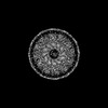
Internal virion protein gp14 / Internal virion protein gp15 / Portal protein / Tail tubular protein gp11 / Tail tubular protein gp12 / Tail fiber protein Similarity search - Component
Biological species
Escherichia phage T7 (virus)
Method
ELECTRON MICROSCOPY / single particle reconstruction / cryo EM / Resolution: 3.2 Å
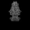
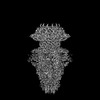
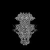
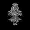
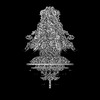
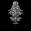
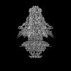
r: Portal protein 0: Tail tubular protein gp11 5: Tail tubular protein gp11 O: Tail fiber protein P: Tail tubular protein gp12 Y: Tail fiber protein a: Internal virion protein gp14 x: Internal virion protein gp15 g: Tail fiber protein u: Portal protein
In the structure databanks used in Yorodumi, some data are registered as the other names, "COVID-19 virus" and "2019-nCoV". Here are the details of the virus and the list of structure data.
Jan 31, 2019. EMDB accession codes are about to change! (news from PDBe EMDB page)
EMDB accession codes are about to change! (news from PDBe EMDB page)
The allocation of 4 digits for EMDB accession codes will soon come to an end. Whilst these codes will remain in use, new EMDB accession codes will include an additional digit and will expand incrementally as the available range of codes is exhausted. The current 4-digit format prefixed with “EMD-” (i.e. EMD-XXXX) will advance to a 5-digit format (i.e. EMD-XXXXX), and so on. It is currently estimated that the 4-digit codes will be depleted around Spring 2019, at which point the 5-digit format will come into force.
The EM Navigator/Yorodumi systems omit the EMD- prefix.
Related info.:Q: What is EMD? / ID/Accession-code notation in Yorodumi/EM Navigator
Yorodumi is a browser for structure data from EMDB, PDB, SASBDB, etc.
This page is also the successor to EM Navigator detail page, and also detail information page/front-end page for Omokage search.
The word "yorodu" (or yorozu) is an old Japanese word meaning "ten thousand". "mi" (miru) is to see.
Related info.:EMDB / PDB / SASBDB / Comparison of 3 databanks / Yorodumi Search / Aug 31, 2016. New EM Navigator & Yorodumi / Yorodumi Papers / Jmol/JSmol / Function and homology information / Changes in new EM Navigator and Yorodumi
 Movie
Movie Controller


 Open data
 Basic information
 Components
 Keywords
 Function and homology information
 Escherichia phage T7 (virus)
 Authors
 United States, 1items
  Citation
 Structure visualization
 Downloads & links
 Other downloads
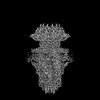
 PDBj
PDBj Assembly
 Sample preparation
 Electron microscopy imaging
 FIELD EMISSION GUN / Accelerating voltage: 300 kV / Illumination mode: FLOOD BEAM
 Processing