 Movie
Movie Controller


+ Open data

- Basic information
| Entry | Database: PDB / ID: 8.0E+41 | ||||||||||||||||||||||||||||||||||||||||||||||||||||||||||||||||||
|---|---|---|---|---|---|---|---|---|---|---|---|---|---|---|---|---|---|---|---|---|---|---|---|---|---|---|---|---|---|---|---|---|---|---|---|---|---|---|---|---|---|---|---|---|---|---|---|---|---|---|---|---|---|---|---|---|---|---|---|---|---|---|---|---|---|---|---|
| Title | E. coli 50S ribosome bound to tiamulin and VS1 | ||||||||||||||||||||||||||||||||||||||||||||||||||||||||||||||||||
 Components Components |
| ||||||||||||||||||||||||||||||||||||||||||||||||||||||||||||||||||
 Keywords Keywords | RIBOSOME / E. coli ribosome / antibiotics | ||||||||||||||||||||||||||||||||||||||||||||||||||||||||||||||||||
| Function / homology |  Function and homology information Function and homology informationDnaA-L2 complex / negative regulation of DNA-templated DNA replication initiation / cytosolic ribosome assembly / ribosome assembly / assembly of large subunit precursor of preribosome / large ribosomal subunit / transferase activity / ribosomal large subunit assembly / large ribosomal subunit rRNA binding / cytosolic large ribosomal subunit ...DnaA-L2 complex / negative regulation of DNA-templated DNA replication initiation / cytosolic ribosome assembly / ribosome assembly / assembly of large subunit precursor of preribosome / large ribosomal subunit / transferase activity / ribosomal large subunit assembly / large ribosomal subunit rRNA binding / cytosolic large ribosomal subunit / cytoplasmic translation / rRNA binding / structural constituent of ribosome / ribosome / translation / ribonucleoprotein complex / response to antibiotic / RNA binding / zinc ion binding / cytoplasm / cytosol Similarity search - Function | ||||||||||||||||||||||||||||||||||||||||||||||||||||||||||||||||||
| Biological species |  Streptomyces virginiae (bacteria) Streptomyces virginiae (bacteria) | ||||||||||||||||||||||||||||||||||||||||||||||||||||||||||||||||||
| Method | ELECTRON MICROSCOPY / single particle reconstruction / cryo EM / Resolution: 2.13 Å | ||||||||||||||||||||||||||||||||||||||||||||||||||||||||||||||||||
 Authors Authors | Pellegrino, J. / Lee, D.J. / Fraser, J.S. / Seiple, I.B. | ||||||||||||||||||||||||||||||||||||||||||||||||||||||||||||||||||
| Funding support |  United States, 3items United States, 3items
| ||||||||||||||||||||||||||||||||||||||||||||||||||||||||||||||||||
 Citation Citation | Journal: To Be Published Title: Tiamulin and NPET binders Authors: Edmonson, Q. / Pellegrino, J. / Lee, D.J. / Seiple, I.B. / Fraser, J.S. | ||||||||||||||||||||||||||||||||||||||||||||||||||||||||||||||||||
| History |
|
- Structure visualization
| Structure viewer | Molecule: MolmilJmol/JSmol |
|---|
- Downloads & links
-Download
| PDBx/mmCIF format | 8e41.cif.gz | 2.4 MB | Display | PDBx/mmCIF format |
|---|---|---|---|---|
| PDB format | pdb8e41.ent.gz | Display | PDB format | |
| PDBx/mmJSON format | 8e41.json.gz | Tree view | PDBx/mmJSON format | |
| Others |  Other downloads Other downloads |
-Validation report
| Arichive directory | https://data.pdbj.org/pub/pdb/validation_reports/e4/8e41ftp://data.pdbj.org/pub/pdb/validation_reports/e4/8e41 | HTTPS FTP |
|---|
-Related structure data
| Related structure data |  27876MC  8e42C M: map data used to model this data C: citing same article ( |
|---|---|
| Similar structure data |
-Links
 PDBj
PDBj


- Assembly
| Deposited unit | 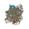
|
|---|---|
| 1 |
|
-Components
-RNA chain , 2 types, 2 molecules IJ
| #1: RNA chain | Mass: 941795.562 Da / Num. of mol.: 1 Source method: isolated from a genetically manipulated source Source: (gene. exp.) |
|---|---|
| #2: RNA chain | Mass: 38177.762 Da / Num. of mol.: 1 Source method: isolated from a genetically manipulated source Source: (gene. exp.) |
-50S ribosomal protein ... , 8 types, 8 molecules KLMNOPQR
| #3: Protein | Mass: 29663.244 Da / Num. of mol.: 1 Source method: isolated from a genetically manipulated source Source: (gene. exp.) |
|---|---|
| #4: Protein | Mass: 15008.471 Da / Num. of mol.: 1 Source method: isolated from a genetically manipulated source Source: (gene. exp.) |
| #5: Protein | Mass: 22121.566 Da / Num. of mol.: 1 Source method: isolated from a genetically manipulated source Source: (gene. exp.) |
| #6: Protein | Mass: 22277.535 Da / Num. of mol.: 1 Source method: isolated from a genetically manipulated source Source: (gene. exp.) |
| #7: Protein | Mass: 16050.606 Da / Num. of mol.: 1 Source method: isolated from a genetically manipulated source Source: (gene. exp.) |
| #8: Protein | Mass: 12253.359 Da / Num. of mol.: 1 Source method: isolated from a genetically manipulated source Source: (gene. exp.) |
| #9: Protein | Mass: 6332.249 Da / Num. of mol.: 1 Source method: isolated from a genetically manipulated source Source: (gene. exp.) |
| #10: Protein/peptide | Mass: 5397.463 Da / Num. of mol.: 1 Source method: isolated from a genetically manipulated source Source: (gene. exp.) |
-Protein/peptide / Non-polymers , 2 types, 2 molecules B
| #11: Protein/peptide |   Type: Cyclic peptide / Class: Antibiotic, Antimicrobial / Mass: 841.907 Da / Num. of mol.: 1 / Source method: isolated from a natural source Type: Cyclic peptide / Class: Antibiotic, Antimicrobial / Mass: 841.907 Da / Num. of mol.: 1 / Source method: isolated from a natural sourceDetails: VIRGINIAMYCIN S1 IS A CYLIC HEPTAPEPTIDE. THE RING IS GENERATED BY LINKING RESIDUE 2 SIDE-CHAIN AND RESIDUE 7 MAIN-CHAIN. Source: (natural) Streptomyces virginiae (bacteria) / References: VIRGINIAMYCIN S1 |
|---|---|
| #12: Chemical | ChemComp-MUL /  Mass: 493.742 Da / Num. of mol.: 1 / Source method: obtained synthetically / Formula: C28H47NO4S / Feature type: SUBJECT OF INVESTIGATION / Comment: antibiotic, Antimicrobial*YM Mass: 493.742 Da / Num. of mol.: 1 / Source method: obtained synthetically / Formula: C28H47NO4S / Feature type: SUBJECT OF INVESTIGATION / Comment: antibiotic, Antimicrobial*YM |
-Details
| Has ligand of interest | Y |
|---|---|
| Has protein modification | Y |
-Experimental details
-Experiment
| Experiment | Method: ELECTRON MICROSCOPY |
|---|---|
| EM experiment | Aggregation state: PARTICLE / 3D reconstruction method: single particle reconstruction |
- Sample preparation
| Component | Name: 50S E. coli ribosome / Type: RIBOSOME / Entity ID: #2-#10 / Source: NATURAL |
|---|---|
| Molecular weight | Experimental value: NO |
| Source (natural) | Organism: |
| Buffer solution | pH: 7.5 |
| Specimen | Embedding applied: NO / Shadowing applied: NO / Staining applied: NO / Vitrification applied: YES |
| Vitrification | Cryogen name: ETHANE |
- Electron microscopy imaging
| Experimental equipment |  Model: Titan Krios / Image courtesy: FEI Company |
|---|---|
| Microscopy | Model: FEI TITAN KRIOS |
| Electron gun | Electron source:  FIELD EMISSION GUN / Accelerating voltage: 300 kV / Illumination mode: FLOOD BEAM FIELD EMISSION GUN / Accelerating voltage: 300 kV / Illumination mode: FLOOD BEAM |
| Electron lens | Mode: BRIGHT FIELD / Nominal defocus max: 1300 nm / Nominal defocus min: 500 nm |
| Image recording | Electron dose: 23.45 e/Å2 / Film or detector model: GATAN K3 BIOQUANTUM (6k x 4k) |
- Processing
| CTF correction | Type: PHASE FLIPPING AND AMPLITUDE CORRECTION |
|---|---|
| 3D reconstruction | Resolution: 2.13 Å / Resolution method: FSC 0.143 CUT-OFF / Num. of particles: 192760 / Symmetry type: POINT |
