 Movie
Movie Controller


+ Open data

- Basic information
| Entry | Database: PDB / ID: 8dyv | |||||||||||||||||||||||||||||||||||||||
|---|---|---|---|---|---|---|---|---|---|---|---|---|---|---|---|---|---|---|---|---|---|---|---|---|---|---|---|---|---|---|---|---|---|---|---|---|---|---|---|---|
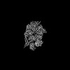
| Title | Structure of human cytoplasmic dynein-1 bound to one Lis1 | |||||||||||||||||||||||||||||||||||||||
 Components Components |
| |||||||||||||||||||||||||||||||||||||||
 Keywords Keywords | MOTOR PROTEIN / dynein / transport | |||||||||||||||||||||||||||||||||||||||
| Function / homology |  Function and homology information Function and homology informationmicrotubule cytoskeleton organization involved in establishment of planar polarity / ameboidal-type cell migration / establishment of planar polarity of embryonic epithelium / 1-alkyl-2-acetylglycerophosphocholine esterase complex / corpus callosum morphogenesis / maintenance of centrosome location / platelet activating factor metabolic process / radial glia-guided pyramidal neuron migration / establishment of centrosome localization / acrosome assembly ...microtubule cytoskeleton organization involved in establishment of planar polarity / ameboidal-type cell migration / establishment of planar polarity of embryonic epithelium / 1-alkyl-2-acetylglycerophosphocholine esterase complex / corpus callosum morphogenesis / maintenance of centrosome location / platelet activating factor metabolic process / radial glia-guided pyramidal neuron migration / establishment of centrosome localization / acrosome assembly / cerebral cortex neuron differentiation / nuclear membrane disassembly / central region of growth cone / microtubule sliding / positive regulation of cytokine-mediated signaling pathway / microtubule organizing center organization / positive regulation of embryonic development / layer formation in cerebral cortex / interneuron migration / astral microtubule / auditory receptor cell development / cortical microtubule organization / myeloid leukocyte migration / reelin-mediated signaling pathway / positive regulation of intracellular transport / regulation of metaphase plate congression / positive regulation of spindle assembly / positive regulation of dendritic spine morphogenesis / osteoclast development / establishment of spindle localization / stereocilium / microtubule plus-end binding / brain morphogenesis / motile cilium / vesicle transport along microtubule / retrograde axonal transport / COPI-independent Golgi-to-ER retrograde traffic / minus-end-directed microtubule motor activity / microtubule associated complex / negative regulation of JNK cascade / P-body assembly / dynein light intermediate chain binding / cytoplasmic dynein complex / kinesin complex / neuromuscular process controlling balance / stem cell division / nuclear migration / establishment of mitotic spindle orientation / dynein intermediate chain binding / cell leading edge / germ cell development / transmission of nerve impulse / dynein complex binding / neuroblast proliferation / dynactin binding / cochlea development / protein secretion / microtubule-based process / positive regulation of axon extension / lipid catabolic process / COPI-mediated anterograde transport / cytoplasmic microtubule / phospholipase binding / JNK cascade / cytoplasmic microtubule organization / Loss of Nlp from mitotic centrosomes / Loss of proteins required for interphase microtubule organization from the centrosome / axon cytoplasm / Amplification of signal from unattached kinetochores via a MAD2 inhibitory signal / Recruitment of mitotic centrosome proteins and complexes / MHC class II antigen presentation / positive regulation of mitotic cell cycle / Recruitment of NuMA to mitotic centrosomes / Anchoring of the basal body to the plasma membrane / HSP90 chaperone cycle for steroid hormone receptors (SHR) in the presence of ligand / Mitotic Prometaphase / EML4 and NUDC in mitotic spindle formation / AURKA Activation by TPX2 / Resolution of Sister Chromatid Cohesion / stress granule assembly / mitotic spindle organization / regulation of microtubule cytoskeleton organization / regulation of mitotic spindle organization / filopodium / adult locomotory behavior / hippocampus development / phosphoprotein binding / RHO GTPases Activate Formins / cerebral cortex development / kinetochore / modulation of chemical synaptic transmission / microtubule cytoskeleton organization / Schaffer collateral - CA1 synapse / neuron migration / HCMV Early Events / Aggrephagy / azurophil granule lumen / Separation of Sister Chromatids / Regulation of PLK1 Activity at G2/M Transition / nuclear envelope Similarity search - Function | |||||||||||||||||||||||||||||||||||||||
| Biological species |  Homo sapiens (human) Homo sapiens (human) | |||||||||||||||||||||||||||||||||||||||
| Method | ELECTRON MICROSCOPY / single particle reconstruction / cryo EM / Resolution: 3.97 Å | |||||||||||||||||||||||||||||||||||||||
 Authors Authors | Reimer, J.M. / DeSantis, M. / Reck-Peterson, S.L. / Leschziner, A.E. | |||||||||||||||||||||||||||||||||||||||
| Funding support |  United States, 2items United States, 2items
| |||||||||||||||||||||||||||||||||||||||
 Citation Citation | Journal: Elife / Year: 2023 Title: Structures of human dynein in complex with the lissencephaly 1 protein, LIS1. Authors: Janice M Reimer / Morgan E DeSantis / Samara L Reck-Peterson / Andres E Leschziner / Abstract: The lissencephaly 1 protein, LIS1, is mutated in type-1 lissencephaly and is a key regulator of cytoplasmic dynein-1. At a molecular level, current models propose that LIS1 activates dynein by ...The lissencephaly 1 protein, LIS1, is mutated in type-1 lissencephaly and is a key regulator of cytoplasmic dynein-1. At a molecular level, current models propose that LIS1 activates dynein by relieving its autoinhibited form. Previously we reported a 3.1 Å structure of yeast dynein bound to Pac1, the yeast homologue of LIS1, which revealed the details of their interactions (Gillies et al., 2022). Based on this structure, we made mutations that disrupted these interactions and showed that they were required for dynein's function in vivo in yeast. We also used our yeast dynein-Pac1 structure to design mutations in human dynein to probe the role of LIS1 in promoting the assembly of active dynein complexes. These mutations had relatively mild effects on dynein activation, suggesting that there may be differences in how dynein and Pac1/LIS1 interact between yeast and humans. Here, we report cryo-EM structures of human dynein-LIS1 complexes. Our new structures reveal the differences between the yeast and human systems, provide a blueprint to disrupt the human dynein-LIS1 interactions more accurately, and map type-1 lissencephaly disease mutations, as well as mutations in dynein linked to malformations of cortical development/intellectual disability, in the context of the dynein-LIS1 complex. #1: Journal: Elife / Year: 2022Title: Structural basis for cytoplasmic dynein-1 regulation by Lis1. Authors: Gillies, J.P. / Reimer, J.M. / Karasmanis, E.P. / Lahiri, I. / Htet, Z.M. / Leschziner, A.E. / Reck-Peterson, S.L. | |||||||||||||||||||||||||||||||||||||||
| History |
|
- Structure visualization
| Structure viewer | Molecule: MolmilJmol/JSmol |
|---|
- Downloads & links
-Download
| PDBx/mmCIF format | 8dyv.cif.gz | 1 MB | Display | PDBx/mmCIF format |
|---|---|---|---|---|
| PDB format | pdb8dyv.ent.gz | 878 KB | Display | PDB format |
| PDBx/mmJSON format | 8dyv.json.gz | Tree view | PDBx/mmJSON format | |
| Others |  Other downloads Other downloads |
-Validation report
| Arichive directory | https://data.pdbj.org/pub/pdb/validation_reports/dy/8dyvftp://data.pdbj.org/pub/pdb/validation_reports/dy/8dyv | HTTPS FTP |
|---|
-Related structure data
| Related structure data | 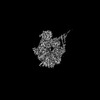 27783MC 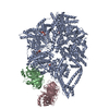 8dyuC M: map data used to model this data C: citing same article ( |
|---|---|
| Similar structure data |
-Links
 PDBj
PDBj


- Assembly
| Deposited unit | 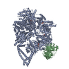
|
|---|---|
| 1 |
|
-Components
| #1: Protein | Mass: 380953.594 Da / Num. of mol.: 1 Source method: isolated from a genetically manipulated source Source: (gene. exp.) Homo sapiens (human) / Production host:   Spodoptera frugiperda (fall armyworm) / References: UniProt: Q14204 Spodoptera frugiperda (fall armyworm) / References: UniProt: Q14204 | ||||||
|---|---|---|---|---|---|---|---|
| #2: Protein | Mass: 46722.918 Da / Num. of mol.: 1 Source method: isolated from a genetically manipulated source Source: (gene. exp.) Homo sapiens (human) / Gene: PAFAH1B1, LIS1, MDCR, MDS, PAFAHA / Production host: Spodoptera frugiperda (fall armyworm) / References: UniProt: P43034 | ||||||
| #3: Chemical |   Mass: 427.201 Da / Num. of mol.: 3 / Source method: obtained synthetically / Formula: C10H15N5O10P2 / Comment: ADP, energy-carrying molecule*YM Mass: 427.201 Da / Num. of mol.: 3 / Source method: obtained synthetically / Formula: C10H15N5O10P2 / Comment: ADP, energy-carrying molecule*YM#4: Chemical | ChemComp-ATP / |   Mass: 507.181 Da / Num. of mol.: 1 / Source method: obtained synthetically / Formula: C10H16N5O13P3 / Comment: ATP, energy-carrying molecule*YM Mass: 507.181 Da / Num. of mol.: 1 / Source method: obtained synthetically / Formula: C10H16N5O13P3 / Comment: ATP, energy-carrying molecule*YMHas ligand of interest | N | Has protein modification | N | |
-Experimental details
-Experiment
| Experiment | Method: ELECTRON MICROSCOPY |
|---|---|
| EM experiment | Aggregation state: PARTICLE / 3D reconstruction method: single particle reconstruction |
- Sample preparation
| Component | Name: Human cytoplasmic dynein-1 bound to one Lis1 / Type: COMPLEX / Entity ID: #1-#2 / Source: RECOMBINANT |
|---|---|
| Source (natural) | Organism: Homo sapiens (human) |
| Source (recombinant) | Organism: Spodoptera frugiperda (fall armyworm) |
| Buffer solution | pH: 7.4 |
| Specimen | Embedding applied: NO / Shadowing applied: NO / Staining applied: NO / Vitrification applied: YES |
| Vitrification | Cryogen name: ETHANE |
- Electron microscopy imaging
| Experimental equipment |  Model: Talos Arctica / Image courtesy: FEI Company |
|---|---|
| Microscopy | Model: FEI TALOS ARCTICA |
| Electron gun | Electron source:  FIELD EMISSION GUN / Accelerating voltage: 200 kV / Illumination mode: FLOOD BEAM FIELD EMISSION GUN / Accelerating voltage: 200 kV / Illumination mode: FLOOD BEAM |
| Electron lens | Mode: BRIGHT FIELD / Nominal defocus max: 2200 nm / Nominal defocus min: 1400 nm |
| Image recording | Electron dose: 55 e/Å2 / Film or detector model: GATAN K2 SUMMIT (4k x 4k) |
- Processing
| Software | Name: PHENIX / Version: 1.20.1_4487: / Classification: refinement | ||||||||||||||||||||||||
|---|---|---|---|---|---|---|---|---|---|---|---|---|---|---|---|---|---|---|---|---|---|---|---|---|---|
| EM software | Name: PHENIX / Category: model refinement | ||||||||||||||||||||||||
| CTF correction | Type: PHASE FLIPPING AND AMPLITUDE CORRECTION | ||||||||||||||||||||||||
| 3D reconstruction | Resolution: 3.97 Å / Resolution method: FSC 0.143 CUT-OFF / Num. of particles: 24417 / Symmetry type: POINT | ||||||||||||||||||||||||
| Refine LS restraints |
|
