 Movie
Movie Controller

[English] 日本語
 Yorodumi
- PDB-8dyk: Room temperature neutron structure of a fluorescent Ag8 cluster t... -
+ Open data

- Basic information
| Entry | Database: PDB / ID: 8dyk | ||||||||||||||||||||||||||||||||||
|---|---|---|---|---|---|---|---|---|---|---|---|---|---|---|---|---|---|---|---|---|---|---|---|---|---|---|---|---|---|---|---|---|---|---|---|
| Title | Room temperature neutron structure of a fluorescent Ag8 cluster templated by a multistranded DNA scaffold | ||||||||||||||||||||||||||||||||||
 Components Components | DNA (5'-D(* Keywords KeywordsDNA / Metal clusters / Silver / chromophore | Function / homology | SILVER ION / DNA |  Function and homology information Function and homology informationBiological species | synthetic construct (others) | Method | NEUTRON DIFFRACTION / |  MOLECULAR REPLACEMENT / Resolution: 2.1 Å MOLECULAR REPLACEMENT / Resolution: 2.1 Å  Authors AuthorsMeilleur, F. / Lieberman, R.L. / Petty, J.T. | Funding support | |  United States, 3items United States, 3items
 CitationJournal: J Phys Chem Lett / Year: 2022 CitationJournal: J Phys Chem Lett / Year: 2022Title: Mapping H + in the Nanoscale (A 2 C 4 ) 2 -Ag 8 Fluorophore. Authors: David, F. / Setzler, C. / Sorescu, A. / Lieberman, R.L. / Meilleur, F. / Petty, J.T. History |
|
- Structure visualization
| Structure viewer | Molecule: MolmilJmol/JSmol |
|---|
- Downloads & links
-Download
| PDBx/mmCIF format | 8dyk.cif.gz | 22.2 KB | Display | PDBx/mmCIF format |
|---|---|---|---|---|
| PDB format | pdb8dyk.ent.gz | 13.5 KB | Display | PDB format |
| PDBx/mmJSON format | 8dyk.json.gz | Tree view | PDBx/mmJSON format | |
| Others |  Other downloads Other downloads |
-Validation report
| Summary document | 8dyk_validation.pdf.gz | 262.3 KB | Display | wwPDB validaton report |
|---|---|---|---|---|
| Full document | 8dyk_full_validation.pdf.gz | 262.2 KB | Display | |
| Data in XML | 8dyk_validation.xml.gz | 1.1 KB | Display | |
| Data in CIF | 8dyk_validation.cif.gz | 1.5 KB | Display | |
| Arichive directory | https://data.pdbj.org/pub/pdb/validation_reports/dy/8dykftp://data.pdbj.org/pub/pdb/validation_reports/dy/8dyk | HTTPS FTP |
-Related structure data
| Related structure data | 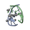 6nizS S: Starting model for refinement |
|---|---|
| Similar structure data |
-Links
 PDBj
PDBj

- Assembly
| Deposited unit | 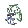
| |||||||||||||||
|---|---|---|---|---|---|---|---|---|---|---|---|---|---|---|---|---|
| 1 |
| |||||||||||||||
| Unit cell |
| |||||||||||||||
| Components on special symmetry positions |
|
-Components
| #1: DNA chain | Mass: 1738.183 Da / Num. of mol.: 2 / Source method: obtained synthetically / Source: (synth.) synthetic construct (others) #2: Chemical | ChemComp-AG /   Mass: 107.868 Da / Num. of mol.: 11 / Source method: obtained synthetically / Formula: Ag / Feature type: SUBJECT OF INVESTIGATION Mass: 107.868 Da / Num. of mol.: 11 / Source method: obtained synthetically / Formula: Ag / Feature type: SUBJECT OF INVESTIGATION#3: Water | ChemComp-HOH / |  Mass: 18.015 Da / Num. of mol.: 24 / Source method: isolated from a natural source / Formula: H2O Mass: 18.015 Da / Num. of mol.: 24 / Source method: isolated from a natural source / Formula: H2OHas ligand of interest | Y | |
|---|
-Experimental details
-Experiment
| Experiment | Method: NEUTRON DIFFRACTION / Number of used crystals: 1 |
|---|
- Sample preparation
| Crystal | Density Matthews: 2.47 Å3/Da / Density % sol: 50.2 % |
|---|---|
| Crystal grow | Temperature: 293 K / Method: vapor diffusion, hanging drop / pH: 6 Details: 1.5 mM A2C4, 9mM AgNO3, 70 mM cacodylate buffer (pH 6), 2-methyl-2,4-pentanediol (MPD) |
-Data collection
| Diffraction | Mean temperature: 293 K / Serial crystal experiment: N | |||||||||
|---|---|---|---|---|---|---|---|---|---|---|
| Diffraction source | Source: SPALLATION SOURCE / Site: ORNL Spallation Neutron Source / Beamline: MANDI / Wavelength: 2.0-4.0 | |||||||||
| Detector | Type: ORNL ANGER CAMERA / Detector: AREA DETECTOR / Date: May 7, 2021 | |||||||||
| Radiation | Protocol: LAUE / Monochromatic (M) / Laue (L): L / Scattering type: neutron | |||||||||
| Radiation wavelength |
| |||||||||
| Reflection | Resolution: 2.1→12.26 Å / Num. obs: 2315 / % possible obs: 96.49 % / Redundancy: 6.2 % / Biso Wilson estimate: 13.1 Å2 / CC1/2: 0.991 / CC star: 0.998 / Rmerge(I) obs: 0.146 / Rpim(I) all: 0.059 / Rrim(I) all: 0.1588 / Net I/σ(I): 10.5 | |||||||||
| Reflection shell | Resolution: 2.1→2.175 Å / Redundancy: 5.6 % / Rmerge(I) obs: 0.2635 / Mean I/σ(I) obs: 3.77 / Num. unique obs: 216 / CC1/2: 0.328 / CC star: 0.703 / Rpim(I) all: 0.1152 / Rrim(I) all: 0.2901 / % possible all: 95.15 |
- Processing
| Software |
| ||||||||||||||||||||||||
|---|---|---|---|---|---|---|---|---|---|---|---|---|---|---|---|---|---|---|---|---|---|---|---|---|---|
| Refinement | Method to determine structure: MOLECULAR REPLACEMENT Starting model: 6NIZ Resolution: 2.1→12.26 Å / SU ML: 0.3736 / Cross valid method: FREE R-VALUE / σ(F): 1.65 / Phase error: 24.9248 Stereochemistry target values: GeoStd + Monomer Library + CDL v1.2
| ||||||||||||||||||||||||
| Solvent computation | Shrinkage radii: 0.9 Å / VDW probe radii: 1.11 Å / Solvent model: FLAT BULK SOLVENT MODEL | ||||||||||||||||||||||||
| Displacement parameters | Biso mean: 18.74 Å2 | ||||||||||||||||||||||||
| Refinement step | Cycle: LAST / Resolution: 2.1→12.26 Å
| ||||||||||||||||||||||||
| Refine LS restraints |
| ||||||||||||||||||||||||
| LS refinement shell |
|
