 Movie
Movie Controller


+ Open data

- Basic information
| Entry | Database: PDB / ID: 8dx0 | ||||||
|---|---|---|---|---|---|---|---|
| Title | VanSC CA domain | ||||||
 Components Components | Histidine kinase | ||||||
 Keywords Keywords | SIGNALING PROTEIN / ATP binding / histidine kinase | ||||||
| Function / homology |  Function and homology information Function and homology informationphosphorelay sensor kinase activity / histidine kinase / metal ion binding / plasma membrane Similarity search - Function | ||||||
| Biological species |  Enterococcus (bacteria) Enterococcus (bacteria) | ||||||
| Method |  X-RAY DIFFRACTION / SYNCHROTRON / MOLECULAR REPLACEMENT / Resolution: 1.45 Å X-RAY DIFFRACTION / SYNCHROTRON / MOLECULAR REPLACEMENT / Resolution: 1.45 Å | ||||||
 Authors Authors | Loll, P.J. | ||||||
| Funding support |  United States, 1items United States, 1items
| ||||||
 Citation Citation | Journal: J.Biol.Chem. / Year: 2023 Title: Structure of VanS from vancomycin-resistant enterococci: A sensor kinase with weak ATP binding. Authors: Grasty, K.C. / Guzik, C. / D'Lauro, E.J. / Padrick, S.B. / Beld, J. / Loll, P.J. | ||||||
| History |
|
- Structure visualization
| Structure viewer | Molecule: MolmilJmol/JSmol |
|---|
- Downloads & links
-Download
| PDBx/mmCIF format | 8dx0.cif.gz | 161.3 KB | Display | PDBx/mmCIF format |
|---|---|---|---|---|
| PDB format | pdb8dx0.ent.gz | 105.3 KB | Display | PDB format |
| PDBx/mmJSON format | 8dx0.json.gz | Tree view | PDBx/mmJSON format | |
| Others |  Other downloads Other downloads |
-Validation report
| Arichive directory | https://data.pdbj.org/pub/pdb/validation_reports/dx/8dx0ftp://data.pdbj.org/pub/pdb/validation_reports/dx/8dx0 | HTTPS FTP |
|---|
-Related structure data


-Links
 PDBj
PDBj

- Assembly
| Deposited unit | 
| ||||||||||||
|---|---|---|---|---|---|---|---|---|---|---|---|---|---|
| 1 | 
| ||||||||||||
| 2 | 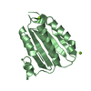
| ||||||||||||
| Unit cell |
|
-Components
| #1: Protein | Mass: 17322.699 Da / Num. of mol.: 2 Source method: isolated from a genetically manipulated source Source: (gene. exp.) Enterococcus (bacteria) / Gene: vanSc / Production host: #2: Chemical |   Mass: 24.305 Da / Num. of mol.: 3 / Source method: obtained synthetically / Formula: Mg Mass: 24.305 Da / Num. of mol.: 3 / Source method: obtained synthetically / Formula: Mg#3: Water | ChemComp-HOH / |  Mass: 18.015 Da / Num. of mol.: 228 / Source method: isolated from a natural source / Formula: H2O Mass: 18.015 Da / Num. of mol.: 228 / Source method: isolated from a natural source / Formula: H2OHas ligand of interest | N | |
|---|
-Experimental details
-Experiment
| Experiment | Method: X-RAY DIFFRACTION / Number of used crystals: 1 |
|---|
- Sample preparation
| Crystal | Density Matthews: 2.35 Å3/Da / Density % sol: 47.76 % |
|---|---|
| Crystal grow | Temperature: 277 K / Method: microbatch / pH: 7 Details: Protein was concentrated to 2.5 mg/mL in 20 mM Tris pH 7.8, 150 mM NaCl supplemented with 2.5 mM AMP-PNP and 5 mM MgCl2. Protein and precipitant were mixed in a volume ratio of 1:3 and ...Details: Protein was concentrated to 2.5 mg/mL in 20 mM Tris pH 7.8, 150 mM NaCl supplemented with 2.5 mM AMP-PNP and 5 mM MgCl2. Protein and precipitant were mixed in a volume ratio of 1:3 and incubated under Als Oil at 277 K; the precipitant solution was 22% (w/v) PEG 6000, 100 mM HEPES buffer at pH 7.0. Crystals of comparable appearance and diffraction quality were obtained using MES buffer at pH 6.0, and Tris buffer at pH 8.0. |
-Data collection
| Diffraction | Mean temperature: 100 K / Serial crystal experiment: N |
|---|---|
| Diffraction source | Source: SYNCHROTRON / Site: NSLS-II / Beamline: 17-ID-1 / Wavelength: 0.9791 Å |
| Detector | Type: DECTRIS EIGER X 9M / Detector: PIXEL / Date: Apr 3, 2019 |
| Radiation | Protocol: SINGLE WAVELENGTH / Monochromatic (M) / Laue (L): M / Scattering type: x-ray |
| Radiation wavelength | Wavelength: 0.9791 Å / Relative weight: 1 |
| Reflection | Resolution: 1.45→64.7 Å / Num. obs: 55912 / % possible obs: 97.1 % / Redundancy: 6.2 % / Biso Wilson estimate: 21.96 Å2 / CC1/2: 0.999 / Rmerge(I) obs: 0.036 / Rpim(I) all: 0.016 / Rrim(I) all: 0.039 / Net I/σ(I): 22.8 |
| Reflection shell | Resolution: 1.45→1.5 Å / Redundancy: 3.1 % / Rmerge(I) obs: 0.6 / Num. unique obs: 4842 / CC1/2: 0.682 / Rpim(I) all: 0.386 / Rrim(I) all: 0.718 / % possible all: 84.7 |
- Processing
| Software |
| ||||||||||||||||||||||||||||||||||||||||||||||||||||||||||||||||||||||||||||||||||||||||||||||||||||||||||||||||||||||||||||||||||||||||||||
|---|---|---|---|---|---|---|---|---|---|---|---|---|---|---|---|---|---|---|---|---|---|---|---|---|---|---|---|---|---|---|---|---|---|---|---|---|---|---|---|---|---|---|---|---|---|---|---|---|---|---|---|---|---|---|---|---|---|---|---|---|---|---|---|---|---|---|---|---|---|---|---|---|---|---|---|---|---|---|---|---|---|---|---|---|---|---|---|---|---|---|---|---|---|---|---|---|---|---|---|---|---|---|---|---|---|---|---|---|---|---|---|---|---|---|---|---|---|---|---|---|---|---|---|---|---|---|---|---|---|---|---|---|---|---|---|---|---|---|---|---|---|
| Refinement | Method to determine structure: MOLECULAR REPLACEMENT Starting model: Six models from SwissModel combined with Ensembler Resolution: 1.45→34.16 Å / SU ML: 0.1662 / Cross valid method: FREE R-VALUE / σ(F): 1.37 / Phase error: 23.1022 / Stereochemistry target values: CDL v1.2
| ||||||||||||||||||||||||||||||||||||||||||||||||||||||||||||||||||||||||||||||||||||||||||||||||||||||||||||||||||||||||||||||||||||||||||||
| Solvent computation | Shrinkage radii: 0.9 Å / VDW probe radii: 1.11 Å / Solvent model: FLAT BULK SOLVENT MODEL | ||||||||||||||||||||||||||||||||||||||||||||||||||||||||||||||||||||||||||||||||||||||||||||||||||||||||||||||||||||||||||||||||||||||||||||
| Displacement parameters | Biso mean: 28.09 Å2 | ||||||||||||||||||||||||||||||||||||||||||||||||||||||||||||||||||||||||||||||||||||||||||||||||||||||||||||||||||||||||||||||||||||||||||||
| Refinement step | Cycle: LAST / Resolution: 1.45→34.16 Å
| ||||||||||||||||||||||||||||||||||||||||||||||||||||||||||||||||||||||||||||||||||||||||||||||||||||||||||||||||||||||||||||||||||||||||||||
| Refine LS restraints |
| ||||||||||||||||||||||||||||||||||||||||||||||||||||||||||||||||||||||||||||||||||||||||||||||||||||||||||||||||||||||||||||||||||||||||||||
| LS refinement shell |
| ||||||||||||||||||||||||||||||||||||||||||||||||||||||||||||||||||||||||||||||||||||||||||||||||||||||||||||||||||||||||||||||||||||||||||||
| Refinement TLS params. | Method: refined / Origin x: 17.04657852 Å / Origin y: 8.49773461621 Å / Origin z: 14.1315729981 Å
| ||||||||||||||||||||||||||||||||||||||||||||||||||||||||||||||||||||||||||||||||||||||||||||||||||||||||||||||||||||||||||||||||||||||||||||
| Refinement TLS group | Selection details: all |
