 Movie
Movie Controller

[English] 日本語
 Yorodumi
- PDB-8dwr: Crystal structure of the L333V variant of catalase-peroxidase fro... -
+ Open data

- Basic information
| Entry | Database: PDB / ID: 8dwr | ||||||
|---|---|---|---|---|---|---|---|
| Title | Crystal structure of the L333V variant of catalase-peroxidase from Mycobacterium tuberculosis | ||||||
 Components Components | Catalase-peroxidase | ||||||
 Keywords Keywords | OXIDOREDUCTASE / Catalase-peroxidase / KatG / Heme | ||||||
| Function / homology |  Function and homology information Function and homology informationoxidoreductase activity, acting on a heme group of donors, nitrogenous group as acceptor / Tolerance of reactive oxygen produced by macrophages / catalase-peroxidase / NADH binding / catalase activity / NADPH binding / peptidoglycan-based cell wall / positive regulation of DNA repair / hydrogen peroxide catabolic process / peroxidase activity ...oxidoreductase activity, acting on a heme group of donors, nitrogenous group as acceptor / Tolerance of reactive oxygen produced by macrophages / catalase-peroxidase / NADH binding / catalase activity / NADPH binding / peptidoglycan-based cell wall / positive regulation of DNA repair / hydrogen peroxide catabolic process / peroxidase activity / cellular response to hydrogen peroxide / response to oxidative stress / response to antibiotic / heme binding / extracellular region / metal ion binding / plasma membrane / cytosol Similarity search - Function | ||||||
| Biological species |   Mycobacterium tuberculosis (bacteria) Mycobacterium tuberculosis (bacteria) | ||||||
| Method |  X-RAY DIFFRACTION / SYNCHROTRON / MOLECULAR REPLACEMENT / Resolution: 2.1 Å X-RAY DIFFRACTION / SYNCHROTRON / MOLECULAR REPLACEMENT / Resolution: 2.1 Å | ||||||
 Authors Authors | Diaz-Vilchis, A. / Uribe-Vazquez, B. / Avila-Linares, A. / Rudino-Pinera, E. / Soberon, X. | ||||||
| Funding support |  Mexico, 1items Mexico, 1items
| ||||||
 Citation Citation | Journal: Biochem Biophys Rep / Year: 2024 Title: Characterization of a catalase-peroxidase variant (L333V-KatG) identified in an INH-resistant Mycobacterium tuberculosis clinical isolate. Authors: Uribe-Vazquez, B. / Diaz-Vilchis, A. / Avila-Linares, A. / Saab-Rincon, G. / Marin-Tovar, Y. / Flores, H. / Pastor, N. / Huerta-Miranda, G. / Rudino-Pinera, E. / Soberon, X. | ||||||
| History |
|
- Structure visualization
| Structure viewer | Molecule: MolmilJmol/JSmol |
|---|
- Downloads & links
-Download
| PDBx/mmCIF format | 8dwr.cif.gz | 601.5 KB | Display | PDBx/mmCIF format |
|---|---|---|---|---|
| PDB format | pdb8dwr.ent.gz | 487.9 KB | Display | PDB format |
| PDBx/mmJSON format | 8dwr.json.gz | Tree view | PDBx/mmJSON format | |
| Others |  Other downloads Other downloads |
-Validation report
| Summary document | 8dwr_validation.pdf.gz | 1.8 MB | Display | wwPDB validaton report |
|---|---|---|---|---|
| Full document | 8dwr_full_validation.pdf.gz | 1.9 MB | Display | |
| Data in XML | 8dwr_validation.xml.gz | 118.6 KB | Display | |
| Data in CIF | 8dwr_validation.cif.gz | 172.6 KB | Display | |
| Arichive directory | https://data.pdbj.org/pub/pdb/validation_reports/dw/8dwrftp://data.pdbj.org/pub/pdb/validation_reports/dw/8dwr | HTTPS FTP |
-Related structure data
| Related structure data | 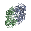 2ccaS S: Starting model for refinement |
|---|---|
| Similar structure data |
-Links
 PDBj
PDBj
- Assembly
| Deposited unit | 
| ||||||||
|---|---|---|---|---|---|---|---|---|---|
| 1 | 
| ||||||||
| 2 | 
| ||||||||
| Unit cell |
|
-Components
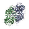
-Protein , 1 types, 4 molecules ABCD
| #1: Protein | Mass: 81534.477 Da / Num. of mol.: 4 / Mutation: L333V Source method: isolated from a genetically manipulated source Source: (gene. exp.) Mycobacterium tuberculosis (bacteria) / Strain: ATCC 25618 / H37Rv / Gene: katG, Rv1908c, MTCY180.10 / Plasmid: pKK-KatG / Production host: |
|---|
-Non-polymers , 5 types, 2209 molecules 


| #2: Chemical | ChemComp-HEM /  Mass: 616.487 Da / Num. of mol.: 4 / Source method: obtained synthetically / Formula: C34H32FeN4O4 / Feature type: SUBJECT OF INVESTIGATION Mass: 616.487 Da / Num. of mol.: 4 / Source method: obtained synthetically / Formula: C34H32FeN4O4 / Feature type: SUBJECT OF INVESTIGATION#3: Chemical | ChemComp-EPE /  Mass: 238.305 Da / Num. of mol.: 4 / Source method: obtained synthetically / Formula: C8H18N2O4S / Comment: pH buffer*YM Mass: 238.305 Da / Num. of mol.: 4 / Source method: obtained synthetically / Formula: C8H18N2O4S / Comment: pH buffer*YM#4: Chemical | ChemComp-NA /  Mass: 22.990 Da / Num. of mol.: 4 / Source method: obtained synthetically / Formula: Na Mass: 22.990 Da / Num. of mol.: 4 / Source method: obtained synthetically / Formula: Na#5: Chemical | ChemComp-PGE /  Mass: 150.173 Da / Num. of mol.: 4 / Source method: obtained synthetically / Formula: C6H14O4 Mass: 150.173 Da / Num. of mol.: 4 / Source method: obtained synthetically / Formula: C6H14O4#6: Water | ChemComp-HOH / | Mass: 18.015 Da / Num. of mol.: 2193 / Source method: isolated from a natural source / Formula: H2O |
|---|
-Details
| Has ligand of interest | Y |
|---|
-Experimental details
-Experiment
| Experiment | Method: X-RAY DIFFRACTION / Number of used crystals: 1 |
|---|
- Sample preparation
| Crystal | Density Matthews: 2.43 Å3/Da / Density % sol: 49.41 % |
|---|---|
| Crystal grow | Temperature: 291 K / Method: vapor diffusion, sitting drop / pH: 7 Details: 10 % w/v PEG 6000 100 mM HEPES / Sodium Hydroxide; pH 7.0 |
-Data collection
| Diffraction | Mean temperature: 100 K / Serial crystal experiment: N |
|---|---|
| Diffraction source | Source: SYNCHROTRON / Site: APS  / Beamline: 19-ID / Wavelength: 0.97918 Å / Beamline: 19-ID / Wavelength: 0.97918 Å |
| Detector | Type: DECTRIS PILATUS 6M / Detector: PIXEL / Date: Jun 13, 2019 |
| Radiation | Protocol: SINGLE WAVELENGTH / Monochromatic (M) / Laue (L): M / Scattering type: x-ray |
| Radiation wavelength | Wavelength: 0.97918 Å / Relative weight: 1 |
| Reflection | Resolution: 2.1→25 Å / Num. obs: 178934 / % possible obs: 98.9 % / Redundancy: 7 % / Biso Wilson estimate: 23 Å2 / CC1/2: 0.99 / Rmerge(I) obs: 0.094 / Net I/σ(I): 9.6 |
| Reflection shell | Resolution: 2.1→2.14 Å / Redundancy: 7.4 % / Rmerge(I) obs: 0.546 / Mean I/σ(I) obs: 3.3 / Num. unique obs: 8861 / CC1/2: 0.8 / % possible all: 99.3 |
- Processing
| Software |
| ||||||||||||||||||||||||||||||||||||||||||||||||||||||||||||||||||||||||||||||||||||||||||
|---|---|---|---|---|---|---|---|---|---|---|---|---|---|---|---|---|---|---|---|---|---|---|---|---|---|---|---|---|---|---|---|---|---|---|---|---|---|---|---|---|---|---|---|---|---|---|---|---|---|---|---|---|---|---|---|---|---|---|---|---|---|---|---|---|---|---|---|---|---|---|---|---|---|---|---|---|---|---|---|---|---|---|---|---|---|---|---|---|---|---|---|
| Refinement | Method to determine structure: MOLECULAR REPLACEMENT Starting model: 2CCA Resolution: 2.1→19.992 Å / SU ML: 0.25 / Cross valid method: FREE R-VALUE / σ(F): 1.33 / Phase error: 22.67 / Stereochemistry target values: ML
| ||||||||||||||||||||||||||||||||||||||||||||||||||||||||||||||||||||||||||||||||||||||||||
| Solvent computation | Shrinkage radii: 0.9 Å / VDW probe radii: 1.11 Å / Solvent model: FLAT BULK SOLVENT MODEL | ||||||||||||||||||||||||||||||||||||||||||||||||||||||||||||||||||||||||||||||||||||||||||
| Displacement parameters | Biso max: 76.3 Å2 / Biso mean: 24.566 Å2 / Biso min: 4.54 Å2 | ||||||||||||||||||||||||||||||||||||||||||||||||||||||||||||||||||||||||||||||||||||||||||
| Refinement step | Cycle: final / Resolution: 2.1→19.992 Å
| ||||||||||||||||||||||||||||||||||||||||||||||||||||||||||||||||||||||||||||||||||||||||||
| Refine LS restraints |
| ||||||||||||||||||||||||||||||||||||||||||||||||||||||||||||||||||||||||||||||||||||||||||
| LS refinement shell | Refine-ID: X-RAY DIFFRACTION / Rfactor Rfree error: 0
|
