 Movie
Movie Controller


+ Open data

- Basic information
| Entry | Database: PDB / ID: 8drj | ||||||
|---|---|---|---|---|---|---|---|
| Title | Apo B2 dimer (H60/H100/H104) formed in the presence of Cu(II) | ||||||
 Components Components | (Soluble cytochrome b562) x 2 | ||||||
 Keywords Keywords | METAL BINDING PROTEIN | ||||||
| Function / homology |  Function and homology information Function and homology informationelectron transport chain / periplasmic space / electron transfer activity / iron ion binding / heme binding Similarity search - Function | ||||||
| Biological species |  | ||||||
| Method |  X-RAY DIFFRACTION / SYNCHROTRON / MOLECULAR REPLACEMENT / Resolution: 2.4 Å X-RAY DIFFRACTION / SYNCHROTRON / MOLECULAR REPLACEMENT / Resolution: 2.4 Å | ||||||
 Authors Authors | Choi, T.S. / Tezcan, F.A. | ||||||
| Funding support |  United States, 1items United States, 1items
| ||||||
 Citation Citation | Journal: J.Am.Chem.Soc. / Year: 2022 Title: Design of a Flexible, Zn-Selective Protein Scaffold that Displays Anti-Irving-Williams Behavior. Authors: Choi, T.S. / Tezcan, F.A. | ||||||
| History |
|
- Structure visualization
| Structure viewer | Molecule: MolmilJmol/JSmol |
|---|
- Downloads & links
-Download
| PDBx/mmCIF format | 8drj.cif.gz | 68.5 KB | Display | PDBx/mmCIF format |
|---|---|---|---|---|
| PDB format | pdb8drj.ent.gz | 40.6 KB | Display | PDB format |
| PDBx/mmJSON format | 8drj.json.gz | Tree view | PDBx/mmJSON format | |
| Others |  Other downloads Other downloads |
-Validation report
| Arichive directory | https://data.pdbj.org/pub/pdb/validation_reports/dr/8drjftp://data.pdbj.org/pub/pdb/validation_reports/dr/8drj | HTTPS FTP |
|---|
-Related structure data
| Related structure data |  8drdC  8drfC  8drlC  8drmC 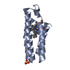 2bc5S S: Starting model for refinement C: citing same article ( |
|---|---|
| Similar structure data |
-Links
 PDBj
PDBj


- Assembly
| Deposited unit | 
| ||||||||||||
|---|---|---|---|---|---|---|---|---|---|---|---|---|---|
| 1 |
| ||||||||||||
| Unit cell |
|
-Components
| #1: Protein | Mass: 11843.324 Da / Num. of mol.: 1 / Mutation: D60H, A100H, K104H Source method: isolated from a genetically manipulated source Source: (gene. exp.) | ||||||||
|---|---|---|---|---|---|---|---|---|---|
| #2: Protein | Mass: 11828.354 Da / Num. of mol.: 1 / Mutation: D60H, A100H, K104H Source method: isolated from a genetically manipulated source Source: (gene. exp.) | ||||||||
| #3: Chemical |   Mass: 618.503 Da / Num. of mol.: 2 / Source method: obtained synthetically / Formula: C34H34FeN4O4 Mass: 618.503 Da / Num. of mol.: 2 / Source method: obtained synthetically / Formula: C34H34FeN4O4#4: Chemical |   Mass: 96.063 Da / Num. of mol.: 2 / Source method: obtained synthetically / Formula: SO4 Mass: 96.063 Da / Num. of mol.: 2 / Source method: obtained synthetically / Formula: SO4#5: Water | ChemComp-HOH / |  Mass: 18.015 Da / Num. of mol.: 4 / Source method: isolated from a natural source / Formula: H2O Mass: 18.015 Da / Num. of mol.: 4 / Source method: isolated from a natural source / Formula: H2OHas ligand of interest | N | Has protein modification | Y | |
-Experimental details
-Experiment
| Experiment | Method: X-RAY DIFFRACTION / Number of used crystals: 1 |
|---|
- Sample preparation
| Crystal | Density Matthews: 2.03 Å3/Da / Density % sol: 39.47 % |
|---|---|
| Crystal grow | Temperature: 298 K / Method: vapor diffusion, sitting drop / pH: 8 Details: PEG3350 25%, (NH4)2SO4 200 mM, Tris 100 mM, CuCl2 4 mM |
-Data collection
| Diffraction | Mean temperature: 100 K / Serial crystal experiment: N |
|---|---|
| Diffraction source | Source: SYNCHROTRON / Site: ALS / Beamline: 8.3.1 / Wavelength: 1.23981 Å |
| Detector | Type: DECTRIS PILATUS 6M / Detector: PIXEL / Date: Oct 16, 2021 |
| Radiation | Protocol: SINGLE WAVELENGTH / Monochromatic (M) / Laue (L): M / Scattering type: x-ray |
| Radiation wavelength | Wavelength: 1.23981 Å / Relative weight: 1 |
| Reflection | Resolution: 2.4→44.86 Å / Num. obs: 7510 / % possible obs: 99.4 % / Redundancy: 1.9 % / Biso Wilson estimate: 23.97 Å2 / CC1/2: 0.996 / CC star: 0.999 / Rmerge(I) obs: 0.03868 / Rpim(I) all: 0.03868 / Rrim(I) all: 0.05471 / Net I/σ(I): 11.32 |
| Reflection shell | Resolution: 2.4→2.486 Å / Redundancy: 1.9 % / Rmerge(I) obs: 0.08279 / Mean I/σ(I) obs: 6.72 / Num. unique obs: 1383 / CC1/2: 0.971 / CC star: 0.993 / Rpim(I) all: 0.08279 / Rrim(I) all: 0.1171 / % possible all: 99.32 |
- Processing
| Software |
| |||||||||||||||||||||||||||||||||||||||||||||||||||||||||||||||||||||||||||||
|---|---|---|---|---|---|---|---|---|---|---|---|---|---|---|---|---|---|---|---|---|---|---|---|---|---|---|---|---|---|---|---|---|---|---|---|---|---|---|---|---|---|---|---|---|---|---|---|---|---|---|---|---|---|---|---|---|---|---|---|---|---|---|---|---|---|---|---|---|---|---|---|---|---|---|---|---|---|---|
| Refinement | Method to determine structure: MOLECULAR REPLACEMENT Starting model: 2bc5 Resolution: 2.4→44.86 Å / SU ML: 0.3436 / Cross valid method: FREE R-VALUE / σ(F): 1.35 / Phase error: 29.7032 Stereochemistry target values: GeoStd + Monomer Library + CDL v1.2
| |||||||||||||||||||||||||||||||||||||||||||||||||||||||||||||||||||||||||||||
| Solvent computation | Shrinkage radii: 0.9 Å / VDW probe radii: 1.11 Å / Solvent model: FLAT BULK SOLVENT MODEL | |||||||||||||||||||||||||||||||||||||||||||||||||||||||||||||||||||||||||||||
| Displacement parameters | Biso mean: 25.66 Å2 | |||||||||||||||||||||||||||||||||||||||||||||||||||||||||||||||||||||||||||||
| Refinement step | Cycle: LAST / Resolution: 2.4→44.86 Å
| |||||||||||||||||||||||||||||||||||||||||||||||||||||||||||||||||||||||||||||
| Refine LS restraints |
| |||||||||||||||||||||||||||||||||||||||||||||||||||||||||||||||||||||||||||||
| LS refinement shell |
|
