 Movie
Movie Controller

[English] 日本語
 Yorodumi
- PDB-8drb: Crystal structure of Neisseria gonorrhoeae carbonic anhydrase wit... -
+ Open data

- Basic information
| Entry | Database: PDB / ID: 8drb | ||||||
|---|---|---|---|---|---|---|---|
| Title | Crystal structure of Neisseria gonorrhoeae carbonic anhydrase with 3-phenyl-N-(5-sulfamoyl-1,3,4-thiadiazol-2-yl)propanamide | ||||||
 Components Components | Carbonic anhydrase | ||||||
 Keywords Keywords | LYASE / Carbonic anhydrase / Neisseria gonorrhoeae / Acetazolamide | ||||||
| Function / homology |  Function and homology information Function and homology informationcarbonic anhydrase / carbonate dehydratase activity / periplasmic space / zinc ion binding Similarity search - Function | ||||||
| Biological species |  Neisseria gonorrhoeae (bacteria) Neisseria gonorrhoeae (bacteria) | ||||||
| Method |  X-RAY DIFFRACTION / SYNCHROTRON / MOLECULAR REPLACEMENT / molecular replacement / Resolution: 2.59 Å X-RAY DIFFRACTION / SYNCHROTRON / MOLECULAR REPLACEMENT / molecular replacement / Resolution: 2.59 Å | ||||||
 Authors Authors | Marapaka, A.K. / Das, C. / Flaherty, D.P. / Yadav, R. | ||||||
| Funding support |  United States, 1items United States, 1items
| ||||||
 Citation Citation | Journal: Acs Med.Chem.Lett. / Year: 2023 Title: Structural Characterization of Thiadiazolesulfonamide Inhibitors Bound to Neisseria gonorrhoeae alpha-Carbonic Anhydrase. Authors: Marapaka, A.K. / Nocentini, A. / Youse, M.S. / An, W. / Holly, K.J. / Das, C. / Yadav, R. / Seleem, M.N. / Supuran, C.T. / Flaherty, D.P. | ||||||
| History |
|
- Structure visualization
| Structure viewer | Molecule: MolmilJmol/JSmol |
|---|
- Downloads & links
-Download
| PDBx/mmCIF format | 8drb.cif.gz | 191 KB | Display | PDBx/mmCIF format |
|---|---|---|---|---|
| PDB format | pdb8drb.ent.gz | 150.8 KB | Display | PDB format |
| PDBx/mmJSON format | 8drb.json.gz | Tree view | PDBx/mmJSON format | |
| Others |  Other downloads Other downloads |
-Validation report
| Arichive directory | https://data.pdbj.org/pub/pdb/validation_reports/dr/8drbftp://data.pdbj.org/pub/pdb/validation_reports/dr/8drb | HTTPS FTP |
|---|
-Related structure data
| Related structure data |  8dpcC  8dqfC  8dr2C  8dyqC  1kopS S: Starting model for refinement C: citing same article ( |
|---|---|
| Similar structure data |
-Links
 PDBj
PDBj
- Assembly
| Deposited unit | 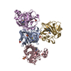
| ||||||||
|---|---|---|---|---|---|---|---|---|---|
| 1 | 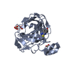
| ||||||||
| 2 | 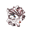
| ||||||||
| 3 | 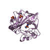
| ||||||||
| 4 | 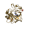
| ||||||||
| Unit cell |
|
-Components
| #1: Protein | Mass: 27315.727 Da / Num. of mol.: 4 Source method: isolated from a genetically manipulated source Source: (gene. exp.) Neisseria gonorrhoeae (bacteria) / Gene: cah / Production host: #2: Chemical | ChemComp-TBW /   Mass: 312.368 Da / Num. of mol.: 4 / Source method: obtained synthetically / Formula: C11H12N4O3S2 / Feature type: SUBJECT OF INVESTIGATION Mass: 312.368 Da / Num. of mol.: 4 / Source method: obtained synthetically / Formula: C11H12N4O3S2 / Feature type: SUBJECT OF INVESTIGATION#3: Chemical | ChemComp-ZN /   Mass: 65.409 Da / Num. of mol.: 4 / Source method: obtained synthetically / Formula: Zn Mass: 65.409 Da / Num. of mol.: 4 / Source method: obtained synthetically / Formula: Zn#4: Chemical | ChemComp-SO4 /   Mass: 96.063 Da / Num. of mol.: 12 / Source method: obtained synthetically / Formula: SO4 Mass: 96.063 Da / Num. of mol.: 12 / Source method: obtained synthetically / Formula: SO4#5: Water | ChemComp-HOH / |  Mass: 18.015 Da / Num. of mol.: 33 / Source method: isolated from a natural source / Formula: H2O Mass: 18.015 Da / Num. of mol.: 33 / Source method: isolated from a natural source / Formula: H2OHas ligand of interest | Y | Has protein modification | Y | |
|---|
-Experimental details
-Experiment
| Experiment | Method: X-RAY DIFFRACTION / Number of used crystals: 1 |
|---|
- Sample preparation
| Crystal | Density Matthews: 2.77 Å3/Da / Density % sol: 55.58 % |
|---|---|
| Crystal grow | Temperature: 298.15 K / Method: vapor diffusion, hanging drop / pH: 7.4 / Details: 0.2 M NH4H2PO4, 2.2 M (NH4)2SO4 / PH range: 6.4-7.4 |
-Data collection
| Diffraction | Mean temperature: 80 K / Serial crystal experiment: N | |||||||||||||||||||||||||||||||||||||||||||||||||||||||||||||||||||||||||||||||||||||||||||||||||||
|---|---|---|---|---|---|---|---|---|---|---|---|---|---|---|---|---|---|---|---|---|---|---|---|---|---|---|---|---|---|---|---|---|---|---|---|---|---|---|---|---|---|---|---|---|---|---|---|---|---|---|---|---|---|---|---|---|---|---|---|---|---|---|---|---|---|---|---|---|---|---|---|---|---|---|---|---|---|---|---|---|---|---|---|---|---|---|---|---|---|---|---|---|---|---|---|---|---|---|---|---|
| Diffraction source | Source: SYNCHROTRON / Site: APS / Beamline: 23-ID-B / Wavelength: 1.0332 Å | |||||||||||||||||||||||||||||||||||||||||||||||||||||||||||||||||||||||||||||||||||||||||||||||||||
| Detector | Type: DECTRIS EIGER X 16M / Detector: PIXEL / Date: Dec 6, 2020 | |||||||||||||||||||||||||||||||||||||||||||||||||||||||||||||||||||||||||||||||||||||||||||||||||||
| Radiation | Protocol: SINGLE WAVELENGTH / Monochromatic (M) / Laue (L): M / Scattering type: x-ray | |||||||||||||||||||||||||||||||||||||||||||||||||||||||||||||||||||||||||||||||||||||||||||||||||||
| Radiation wavelength | Wavelength: 1.0332 Å / Relative weight: 1 | |||||||||||||||||||||||||||||||||||||||||||||||||||||||||||||||||||||||||||||||||||||||||||||||||||
| Reflection | Resolution: 2.59→50 Å / Num. obs: 34669 / % possible obs: 94.1 % / Redundancy: 5.1 % / Rmerge(I) obs: 0.164 / Rpim(I) all: 0.076 / Rrim(I) all: 0.181 / Χ2: 1.03 / Net I/σ(I): 4.9 / Num. measured all: 176488 | |||||||||||||||||||||||||||||||||||||||||||||||||||||||||||||||||||||||||||||||||||||||||||||||||||
| Reflection shell | Diffraction-ID: 1
|
-Phasing
| Phasing | Method: molecular replacement | ||||||
|---|---|---|---|---|---|---|---|
| Phasing MR | R rigid body: 0.547
|
- Processing
| Software |
| |||||||||||||||||||||||||||||||||||||||||||||||||||||||
|---|---|---|---|---|---|---|---|---|---|---|---|---|---|---|---|---|---|---|---|---|---|---|---|---|---|---|---|---|---|---|---|---|---|---|---|---|---|---|---|---|---|---|---|---|---|---|---|---|---|---|---|---|---|---|---|---|
| Refinement | Method to determine structure: MOLECULAR REPLACEMENT Starting model: 1KOP Resolution: 2.59→46.03 Å / Cor.coef. Fo:Fc: 0.938 / Cor.coef. Fo:Fc free: 0.902 / SU B: 10.571 / SU ML: 0.227 / SU R Cruickshank DPI: 0.6691 / Cross valid method: THROUGHOUT / σ(F): 0 / ESU R: 0.669 / ESU R Free: 0.318 / Stereochemistry target values: MAXIMUM LIKELIHOOD Details: HYDROGENS HAVE BEEN ADDED IN THE RIDING POSITIONS U VALUES : REFINED INDIVIDUALLY
| |||||||||||||||||||||||||||||||||||||||||||||||||||||||
| Solvent computation | Ion probe radii: 0.8 Å / Shrinkage radii: 0.8 Å / VDW probe radii: 1.2 Å / Solvent model: MASK | |||||||||||||||||||||||||||||||||||||||||||||||||||||||
| Displacement parameters | Biso max: 175.02 Å2 / Biso mean: 43.375 Å2 / Biso min: 11.22 Å2
| |||||||||||||||||||||||||||||||||||||||||||||||||||||||
| Refinement step | Cycle: final / Resolution: 2.59→46.03 Å
| |||||||||||||||||||||||||||||||||||||||||||||||||||||||
| Refine LS restraints |
| |||||||||||||||||||||||||||||||||||||||||||||||||||||||
| LS refinement shell | Resolution: 2.591→2.658 Å / Rfactor Rfree error: 0 / Total num. of bins used: 20
|
