 Movie
Movie Controller

[English] 日本語
 Yorodumi
- PDB-8dqj: Crystal structure of pyrrolysyl-tRNA synthetase from Methanomethy... -
+ Open data

- Basic information
| Entry | Database: PDB / ID: 8dqj | |||||||||
|---|---|---|---|---|---|---|---|---|---|---|
| Title | Crystal structure of pyrrolysyl-tRNA synthetase from Methanomethylophilus alvus engineered for acridone amino acid (AST) bound to ATP and acridone | |||||||||
 Components Components | AA_TRNA_LIGASE_II domain-containing protein | |||||||||
 Keywords Keywords | LIGASE / Genetic code expansion / Methanomethylophilus alvus / pyrrolysyl-tRNA synthetase / acridone | |||||||||
| Function / homology |  Function and homology information Function and homology information | |||||||||
| Biological species |  Candidatus Methanomethylophilus alvus (archaea) Candidatus Methanomethylophilus alvus (archaea) | |||||||||
| Method |  X-RAY DIFFRACTION / SYNCHROTRON / MOLECULAR REPLACEMENT / Resolution: 1.54 Å X-RAY DIFFRACTION / SYNCHROTRON / MOLECULAR REPLACEMENT / Resolution: 1.54 Å | |||||||||
 Authors Authors | Gottfried-Lee, I. / Karplus, P.A. / Mehl, R.A. / Cooley, R.B. | |||||||||
| Funding support |  United States, 2items United States, 2items
| |||||||||
 Citation Citation | Journal: Acs Chem.Biol. / Year: 2022 Title: Structures of Methanomethylophilus alvus Pyrrolysine tRNA-Synthetases Support the Need for De Novo Selections When Altering the Substrate Specificity. Authors: Gottfried-Lee, I. / Perona, J.J. / Karplus, P.A. / Mehl, R.A. / Cooley, R.B. | |||||||||
| History |
|
- Structure visualization
| Structure viewer | Molecule: MolmilJmol/JSmol |
|---|
- Downloads & links
-Download
| PDBx/mmCIF format | 8dqj.cif.gz | 316.4 KB | Display | PDBx/mmCIF format |
|---|---|---|---|---|
| PDB format | pdb8dqj.ent.gz | 259.8 KB | Display | PDB format |
| PDBx/mmJSON format | 8dqj.json.gz | Tree view | PDBx/mmJSON format | |
| Others |  Other downloads Other downloads |
-Validation report
| Summary document | 8dqj_validation.pdf.gz | 1.5 MB | Display | wwPDB validaton report |
|---|---|---|---|---|
| Full document | 8dqj_full_validation.pdf.gz | 1.5 MB | Display | |
| Data in XML | 8dqj_validation.xml.gz | 25.4 KB | Display | |
| Data in CIF | 8dqj_validation.cif.gz | 37.1 KB | Display | |
| Arichive directory | https://data.pdbj.org/pub/pdb/validation_reports/dq/8dqjftp://data.pdbj.org/pub/pdb/validation_reports/dq/8dqj | HTTPS FTP |
-Related structure data
| Related structure data | 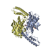 8dqgC 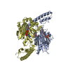 8dqhC 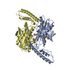 8dqiC 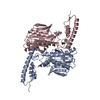 6jp2S C: citing same article ( S: Starting model for refinement |
|---|---|
| Similar structure data |
-Links
 PDBj
PDBj


- Assembly
| Deposited unit | 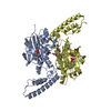
| ||||||||||
|---|---|---|---|---|---|---|---|---|---|---|---|
| 1 |
| ||||||||||
| Unit cell |
|
-Components
-Protein , 1 types, 2 molecules DA
| #1: Protein | Mass: 30750.914 Da / Num. of mol.: 2 / Mutation: N166S V168C W239T Source method: isolated from a genetically manipulated source Source: (gene. exp.) Candidatus Methanomethylophilus alvus (archaea)Gene: BKD89_05515 / Production host:  |
|---|
-Non-polymers , 7 types, 382 molecules 


| #2: Chemical | ChemComp-ATP /  Mass: 507.181 Da / Num. of mol.: 1 / Source method: obtained synthetically / Formula: C10H16N5O13P3 / Comment: ATP, energy-carrying molecule*YM Mass: 507.181 Da / Num. of mol.: 1 / Source method: obtained synthetically / Formula: C10H16N5O13P3 / Comment: ATP, energy-carrying molecule*YM | ||||||||
|---|---|---|---|---|---|---|---|---|---|
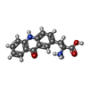
| #3: Chemical | ChemComp-T7Q / ( Type: peptide linking / Mass: 283.302 Da / Num. of mol.: 1 / Source method: obtained synthetically / Formula: C16H15N2O3 / Feature type: SUBJECT OF INVESTIGATION Type: peptide linking / Mass: 283.302 Da / Num. of mol.: 1 / Source method: obtained synthetically / Formula: C16H15N2O3 / Feature type: SUBJECT OF INVESTIGATION | ||||||||
| #4: Chemical |  Mass: 24.305 Da / Num. of mol.: 3 / Source method: obtained synthetically / Formula: Mg Mass: 24.305 Da / Num. of mol.: 3 / Source method: obtained synthetically / Formula: Mg#5: Chemical | ChemComp-GOL / |  Mass: 92.094 Da / Num. of mol.: 1 / Source method: obtained synthetically / Formula: C3H8O3 Mass: 92.094 Da / Num. of mol.: 1 / Source method: obtained synthetically / Formula: C3H8O3#6: Chemical | ChemComp-AMP / |  Mass: 347.221 Da / Num. of mol.: 1 / Source method: obtained synthetically / Formula: C10H14N5O7P / Feature type: SUBJECT OF INVESTIGATION / Comment: AMP*YM Mass: 347.221 Da / Num. of mol.: 1 / Source method: obtained synthetically / Formula: C10H14N5O7P / Feature type: SUBJECT OF INVESTIGATION / Comment: AMP*YM#7: Chemical | ChemComp-PEG / |  Mass: 106.120 Da / Num. of mol.: 1 / Source method: obtained synthetically / Formula: C4H10O3 Mass: 106.120 Da / Num. of mol.: 1 / Source method: obtained synthetically / Formula: C4H10O3#8: Water | ChemComp-HOH / | Mass: 18.015 Da / Num. of mol.: 374 / Source method: isolated from a natural source / Formula: H2O |
-Details
| Has ligand of interest | Y |
|---|
-Experimental details
-Experiment
| Experiment | Method: X-RAY DIFFRACTION / Number of used crystals: 1 |
|---|
- Sample preparation
| Crystal | Density Matthews: 2.84 Å3/Da / Density % sol: 56.76 % |
|---|---|
| Crystal grow | Temperature: 298 K / Method: vapor diffusion, hanging drop / Details: PEG 3350, Sodium citrate tribasic dihydrate |
-Data collection
| Diffraction | Mean temperature: 100 K / Serial crystal experiment: N | ||||||||||||||||||||||||||||||
|---|---|---|---|---|---|---|---|---|---|---|---|---|---|---|---|---|---|---|---|---|---|---|---|---|---|---|---|---|---|---|---|
| Diffraction source | Source: SYNCHROTRON / Site: ALS / Beamline: 5.0.3 / Wavelength: 0.976 Å | ||||||||||||||||||||||||||||||
| Detector | Type: ADSC QUANTUM 315 / Detector: CCD / Date: Oct 29, 2021 | ||||||||||||||||||||||||||||||
| Radiation | Protocol: SINGLE WAVELENGTH / Monochromatic (M) / Laue (L): M / Scattering type: x-ray | ||||||||||||||||||||||||||||||
| Radiation wavelength | Wavelength: 0.976 Å / Relative weight: 1 | ||||||||||||||||||||||||||||||
| Reflection | Resolution: 1.54→35.12 Å / Num. obs: 101330 / % possible obs: 100 % / Redundancy: 13 % / Biso Wilson estimate: 25.97 Å2 / CC1/2: 0.999 / Rmerge(I) obs: 0.099 / Rpim(I) all: 0.029 / Rrim(I) all: 0.103 / Net I/σ(I): 10.6 / Num. measured all: 1312985 / Scaling rejects: 187 | ||||||||||||||||||||||||||||||
| Reflection shell | Diffraction-ID: 1
|
- Processing
| Software |
| ||||||||||||||||||||||||||||||||||||||||||||||||||||||||||||||||||||||||||||||||||||||||||||||||||||||||||||||||||||||||||||||||||||||||||||||||||||||||||||||||||||||||||||||||||||||||||
|---|---|---|---|---|---|---|---|---|---|---|---|---|---|---|---|---|---|---|---|---|---|---|---|---|---|---|---|---|---|---|---|---|---|---|---|---|---|---|---|---|---|---|---|---|---|---|---|---|---|---|---|---|---|---|---|---|---|---|---|---|---|---|---|---|---|---|---|---|---|---|---|---|---|---|---|---|---|---|---|---|---|---|---|---|---|---|---|---|---|---|---|---|---|---|---|---|---|---|---|---|---|---|---|---|---|---|---|---|---|---|---|---|---|---|---|---|---|---|---|---|---|---|---|---|---|---|---|---|---|---|---|---|---|---|---|---|---|---|---|---|---|---|---|---|---|---|---|---|---|---|---|---|---|---|---|---|---|---|---|---|---|---|---|---|---|---|---|---|---|---|---|---|---|---|---|---|---|---|---|---|---|---|---|---|---|---|---|
| Refinement | Method to determine structure: MOLECULAR REPLACEMENT Starting model: 6JP2 Resolution: 1.54→35.12 Å / SU ML: 0.19 / Cross valid method: THROUGHOUT / σ(F): 1.33 / Phase error: 20.76 / Stereochemistry target values: ML
| ||||||||||||||||||||||||||||||||||||||||||||||||||||||||||||||||||||||||||||||||||||||||||||||||||||||||||||||||||||||||||||||||||||||||||||||||||||||||||||||||||||||||||||||||||||||||||
| Solvent computation | Shrinkage radii: 0.9 Å / VDW probe radii: 1.1 Å / Solvent model: FLAT BULK SOLVENT MODEL | ||||||||||||||||||||||||||||||||||||||||||||||||||||||||||||||||||||||||||||||||||||||||||||||||||||||||||||||||||||||||||||||||||||||||||||||||||||||||||||||||||||||||||||||||||||||||||
| Displacement parameters | Biso max: 111.76 Å2 / Biso mean: 39.8651 Å2 / Biso min: 17.69 Å2 | ||||||||||||||||||||||||||||||||||||||||||||||||||||||||||||||||||||||||||||||||||||||||||||||||||||||||||||||||||||||||||||||||||||||||||||||||||||||||||||||||||||||||||||||||||||||||||
| Refinement step | Cycle: final / Resolution: 1.54→35.12 Å
| ||||||||||||||||||||||||||||||||||||||||||||||||||||||||||||||||||||||||||||||||||||||||||||||||||||||||||||||||||||||||||||||||||||||||||||||||||||||||||||||||||||||||||||||||||||||||||
| LS refinement shell | Refine-ID: X-RAY DIFFRACTION / Rfactor Rfree error: 0 / Total num. of bins used: 30 / % reflection obs: 100 %
|
