 Movie
Movie Controller

[English] 日本語
 Yorodumi
- PDB-8dqb: Crystal structure of 3-dehydroquinate dehydratase I from Klebsiel... -
+ Open data

- Basic information
| Entry | Database: PDB / ID: 8dqb | |||||||||
|---|---|---|---|---|---|---|---|---|---|---|
| Title | Crystal structure of 3-dehydroquinate dehydratase I from Klebsiella oxytoca (I23 Form) | |||||||||
 Components Components | 3-dehydroquinate dehydratase I | |||||||||
 Keywords Keywords | HYDROLASE / SSGCID / STRUCTURAL GENOMICS / SEATTLE STRUCTURAL GENOMICS CENTER FOR INFECTIOUS DISEASE | |||||||||
| Function / homology |  Function and homology information Function and homology information5-amino-6-(5-phosphoribosylamino)uracil reductase / diaminohydroxyphosphoribosylaminopyrimidine deaminase Similarity search - Function | |||||||||
| Biological species |  Klebsiella oxytoca (bacteria) Klebsiella oxytoca (bacteria) | |||||||||
| Method |  X-RAY DIFFRACTION / SYNCHROTRON / MOLECULAR REPLACEMENT / molecular replacement / Resolution: 2.5 Å X-RAY DIFFRACTION / SYNCHROTRON / MOLECULAR REPLACEMENT / molecular replacement / Resolution: 2.5 Å | |||||||||
 Authors Authors | Seattle Structural Genomics Center for Infectious Disease / Seattle Structural Genomics Center for Infectious Disease (SSGCID) | |||||||||
| Funding support |  United States, 2items United States, 2items
| |||||||||
 Citation Citation | Journal: To be published Title: Crystal structure of 3-dehydroquinate dehydratase I from Klebsiella oxytoca (I23 Form) Authors: Liu, L. / Seibold, S. / Battaile, K.P. / Lovell, S. | |||||||||
| History |
|
- Structure visualization
| Structure viewer | Molecule: MolmilJmol/JSmol |
|---|
- Downloads & links
-Download
| PDBx/mmCIF format | 8dqb.cif.gz | 157.9 KB | Display | PDBx/mmCIF format |
|---|---|---|---|---|
| PDB format | pdb8dqb.ent.gz | 123.6 KB | Display | PDB format |
| PDBx/mmJSON format | 8dqb.json.gz | Tree view | PDBx/mmJSON format | |
| Others |  Other downloads Other downloads |
-Validation report
| Arichive directory | https://data.pdbj.org/pub/pdb/validation_reports/dq/8dqbftp://data.pdbj.org/pub/pdb/validation_reports/dq/8dqb | HTTPS FTP |
|---|
-Related structure data
| Related structure data | 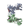 2o7pS S: Starting model for refinement |
|---|---|
| Similar structure data |
-Links
 PDBj
PDBj
- Assembly
| Deposited unit | 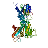
| ||||||||
|---|---|---|---|---|---|---|---|---|---|
| 1 | 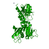
| ||||||||
| Unit cell |
|
-Components
| #1: Protein | Mass: 41256.199 Da / Num. of mol.: 1 / Fragment: KloxA.17380.a.B1 Source method: isolated from a genetically manipulated source Source: (gene. exp.) Klebsiella oxytoca (bacteria) / Plasmid: KloxA.17380.a.B1 / Production host: | ||||
|---|---|---|---|---|---|
| #2: Chemical |   Mass: 65.409 Da / Num. of mol.: 2 / Source method: obtained synthetically / Formula: Zn / Feature type: SUBJECT OF INVESTIGATION Mass: 65.409 Da / Num. of mol.: 2 / Source method: obtained synthetically / Formula: Zn / Feature type: SUBJECT OF INVESTIGATION#3: Water | ChemComp-HOH / |  Mass: 18.015 Da / Num. of mol.: 31 / Source method: isolated from a natural source / Formula: H2O Mass: 18.015 Da / Num. of mol.: 31 / Source method: isolated from a natural source / Formula: H2OHas ligand of interest | Y | |
-Experimental details
-Experiment
| Experiment | Method: X-RAY DIFFRACTION / Number of used crystals: 1 |
|---|
- Sample preparation
| Crystal | Density Matthews: 4.46 Å3/Da / Density % sol: 72.45 % |
|---|---|
| Crystal grow | Temperature: 291 K / Method: vapor diffusion, sitting drop / pH: 6.5 Details: Morpheus A2: 20%(v/v) Ethylene glycol, 10%(w/v) PEG 8000, 100 mM Imidazole/MES, pH 6.5, 30 mM MgCl2 and 30 mM CaCl2,KloxA.17380.a.B1.PW39095 at 5 mg/mL with 2.5 GDP added, no electron ...Details: Morpheus A2: 20%(v/v) Ethylene glycol, 10%(w/v) PEG 8000, 100 mM Imidazole/MES, pH 6.5, 30 mM MgCl2 and 30 mM CaCl2,KloxA.17380.a.B1.PW39095 at 5 mg/mL with 2.5 GDP added, no electron density observed for the GDP, Tray: plate 12679, well A2 drop 3, Puck: PSL1405, Cryo: DIRECT |
-Data collection
| Diffraction | Mean temperature: 100 K / Serial crystal experiment: N | ||||||||||||||||||||||||||||||
|---|---|---|---|---|---|---|---|---|---|---|---|---|---|---|---|---|---|---|---|---|---|---|---|---|---|---|---|---|---|---|---|
| Diffraction source | Source: SYNCHROTRON / Site: NSLS-II / Beamline: 19-ID / Wavelength: 0.97856 Å | ||||||||||||||||||||||||||||||
| Detector | Type: DECTRIS EIGER2 XE 9M / Detector: PIXEL / Date: Jun 7, 2022 | ||||||||||||||||||||||||||||||
| Radiation | Monochromator: Double Crystal Si 111 / Protocol: SINGLE WAVELENGTH / Monochromatic (M) / Laue (L): M / Scattering type: x-ray | ||||||||||||||||||||||||||||||
| Radiation wavelength | Wavelength: 0.97856 Å / Relative weight: 1 | ||||||||||||||||||||||||||||||
| Reflection | Resolution: 2.5→116.05 Å / Num. obs: 25537 / % possible obs: 100 % / Redundancy: 10.4 % / Biso Wilson estimate: 56.22 Å2 / CC1/2: 0.999 / Rmerge(I) obs: 0.089 / Rpim(I) all: 0.029 / Rrim(I) all: 0.094 / Net I/σ(I): 18.6 / Num. measured all: 264742 / Scaling rejects: 42 | ||||||||||||||||||||||||||||||
| Reflection shell | Diffraction-ID: 1
|
-Phasing
| Phasing | Method: molecular replacement |
|---|
- Processing
| Software |
| ||||||||||||||||||||||||||||||||||||||||||||||||||||||||||||||||||||||||||||||||||||||||||||||||||||||||||||||||||||||||||||||||||||||||||||||||||||||
|---|---|---|---|---|---|---|---|---|---|---|---|---|---|---|---|---|---|---|---|---|---|---|---|---|---|---|---|---|---|---|---|---|---|---|---|---|---|---|---|---|---|---|---|---|---|---|---|---|---|---|---|---|---|---|---|---|---|---|---|---|---|---|---|---|---|---|---|---|---|---|---|---|---|---|---|---|---|---|---|---|---|---|---|---|---|---|---|---|---|---|---|---|---|---|---|---|---|---|---|---|---|---|---|---|---|---|---|---|---|---|---|---|---|---|---|---|---|---|---|---|---|---|---|---|---|---|---|---|---|---|---|---|---|---|---|---|---|---|---|---|---|---|---|---|---|---|---|---|---|---|---|
| Refinement | Method to determine structure: MOLECULAR REPLACEMENT Starting model: 2O7P Resolution: 2.5→41.03 Å / SU ML: 0.35 / Cross valid method: THROUGHOUT / σ(F): 1.34 / Phase error: 26.45 / Stereochemistry target values: ML
| ||||||||||||||||||||||||||||||||||||||||||||||||||||||||||||||||||||||||||||||||||||||||||||||||||||||||||||||||||||||||||||||||||||||||||||||||||||||
| Solvent computation | Shrinkage radii: 0.9 Å / VDW probe radii: 1.1 Å / Solvent model: FLAT BULK SOLVENT MODEL | ||||||||||||||||||||||||||||||||||||||||||||||||||||||||||||||||||||||||||||||||||||||||||||||||||||||||||||||||||||||||||||||||||||||||||||||||||||||
| Displacement parameters | Biso max: 114.86 Å2 / Biso mean: 58.4844 Å2 / Biso min: 34.46 Å2 | ||||||||||||||||||||||||||||||||||||||||||||||||||||||||||||||||||||||||||||||||||||||||||||||||||||||||||||||||||||||||||||||||||||||||||||||||||||||
| Refinement step | Cycle: final / Resolution: 2.5→41.03 Å
| ||||||||||||||||||||||||||||||||||||||||||||||||||||||||||||||||||||||||||||||||||||||||||||||||||||||||||||||||||||||||||||||||||||||||||||||||||||||
| LS refinement shell | Refine-ID: X-RAY DIFFRACTION / Rfactor Rfree error: 0 / Total num. of bins used: 9
| ||||||||||||||||||||||||||||||||||||||||||||||||||||||||||||||||||||||||||||||||||||||||||||||||||||||||||||||||||||||||||||||||||||||||||||||||||||||
| Refinement TLS params. | Method: refined / Refine-ID: X-RAY DIFFRACTION
| ||||||||||||||||||||||||||||||||||||||||||||||||||||||||||||||||||||||||||||||||||||||||||||||||||||||||||||||||||||||||||||||||||||||||||||||||||||||
| Refinement TLS group |
|
