 Movie
Movie Controller

[English] 日本語
 Yorodumi
- PDB-8dq2: X-ray crystal structure of Hansschlegelia quercus lanmodulin (Lan... -
+ Open data

- Basic information
| Entry | Database: PDB / ID: 8dq2 | ||||||
|---|---|---|---|---|---|---|---|
| Title | X-ray crystal structure of Hansschlegelia quercus lanmodulin (LanM) with lanthanum (III) bound at pH 7 | ||||||
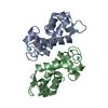
 Components Components | EF-hand domain-containing protein | ||||||
 Keywords Keywords | METAL BINDING PROTEIN / methanol dehydrogenase | ||||||
| Function / homology | EF-Hand 1, calcium-binding site / EF-hand calcium-binding domain. / EF-hand calcium-binding domain profile. / EF-hand domain / EF-hand domain pair / calcium ion binding / CITRIC ACID / LANTHANUM (III) ION / EF-hand domain-containing protein Function and homology information Function and homology information | ||||||
| Biological species |  Hansschlegelia quercus (bacteria) Hansschlegelia quercus (bacteria) | ||||||
| Method |  X-RAY DIFFRACTION / SYNCHROTRON / SAD / Resolution: 1.8 Å X-RAY DIFFRACTION / SYNCHROTRON / SAD / Resolution: 1.8 Å | ||||||
 Authors Authors | Jung, J.J. / Lin, C.-Y. / Boal, A.K. | ||||||
| Funding support |  United States, 1items United States, 1items
| ||||||
 Citation Citation | Journal: Nature / Year: 2023 Title: Enhanced rare-earth separation with a metal-sensitive lanmodulin dimer. Authors: Mattocks, J.A. / Jung, J.J. / Lin, C.Y. / Dong, Z. / Yennawar, N.H. / Featherston, E.R. / Kang-Yun, C.S. / Hamilton, T.A. / Park, D.M. / Boal, A.K. / Cotruvo Jr., J.A. | ||||||
| History |
|
- Structure visualization
| Structure viewer | Molecule: MolmilJmol/JSmol |
|---|
- Downloads & links
-Download
| PDBx/mmCIF format | 8dq2.cif.gz | 109.1 KB | Display | PDBx/mmCIF format |
|---|---|---|---|---|
| PDB format | pdb8dq2.ent.gz | 81.3 KB | Display | PDB format |
| PDBx/mmJSON format | 8dq2.json.gz | Tree view | PDBx/mmJSON format | |
| Others |  Other downloads Other downloads |
-Validation report
| Arichive directory | https://data.pdbj.org/pub/pdb/validation_reports/dq/8dq2ftp://data.pdbj.org/pub/pdb/validation_reports/dq/8dq2 | HTTPS FTP |
|---|
-Related structure data

-Links
 PDBj
PDBj- Assembly
| Deposited unit | 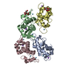
| ||||||||
|---|---|---|---|---|---|---|---|---|---|
| 1 | 
| ||||||||
| 2 | 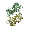
| ||||||||
| Unit cell |
|
-Components
| #1: Protein | Mass: 11923.299 Da / Num. of mol.: 4 Source method: isolated from a genetically manipulated source Source: (gene. exp.) Hansschlegelia quercus (bacteria) / Gene: EYR15_14300 / Production host: #2: Chemical | ChemComp-LA /   Mass: 138.905 Da / Num. of mol.: 12 / Source method: obtained synthetically / Formula: La / Feature type: SUBJECT OF INVESTIGATION Mass: 138.905 Da / Num. of mol.: 12 / Source method: obtained synthetically / Formula: La / Feature type: SUBJECT OF INVESTIGATION#3: Chemical | ChemComp-NA /   Mass: 22.990 Da / Num. of mol.: 4 / Source method: obtained synthetically / Formula: Na Mass: 22.990 Da / Num. of mol.: 4 / Source method: obtained synthetically / Formula: Na#4: Chemical |   Mass: 192.124 Da / Num. of mol.: 2 / Source method: obtained synthetically / Formula: C6H8O7 Mass: 192.124 Da / Num. of mol.: 2 / Source method: obtained synthetically / Formula: C6H8O7#5: Water | ChemComp-HOH / |  Mass: 18.015 Da / Num. of mol.: 263 / Source method: isolated from a natural source / Formula: H2O Mass: 18.015 Da / Num. of mol.: 263 / Source method: isolated from a natural source / Formula: H2OHas ligand of interest | Y | |
|---|
-Experimental details
-Experiment
| Experiment | Method: X-RAY DIFFRACTION / Number of used crystals: 1 |
|---|
- Sample preparation
| Crystal | Density Matthews: 2.04 Å3/Da / Density % sol: 39.77 % |
|---|---|
| Crystal grow | Temperature: 294 K / Method: vapor diffusion, sitting drop / pH: 7 / Details: 0.01 M tri-sodium citrate, 27 % (w/v) PEG 6000 |
-Data collection
| Diffraction | Mean temperature: 100 K / Serial crystal experiment: N |
|---|---|
| Diffraction source | Source: SYNCHROTRON / Site: APS / Beamline: 21-ID-G / Wavelength: 0.97857 Å |
| Detector | Type: MARMOSAIC 300 mm CCD / Detector: CCD / Date: Mar 29, 2022 |
| Radiation | Protocol: SINGLE WAVELENGTH / Monochromatic (M) / Laue (L): M / Scattering type: x-ray |
| Radiation wavelength | Wavelength: 0.97857 Å / Relative weight: 1 |
| Reflection | Resolution: 1.8→50 Å / Num. obs: 41911 / % possible obs: 86.7 % / Redundancy: 6 % / Biso Wilson estimate: 17.53 Å2 / CC1/2: 0.995 / CC star: 0.999 / Rpim(I) all: 0.052 / Rrim(I) all: 0.133 / Net I/σ(I): 12.9 |
| Reflection shell | Resolution: 1.8→1.865 Å / Redundancy: 4.2 % / Mean I/σ(I) obs: 1.39 / Num. unique obs: 1053 / CC1/2: 0.612 / CC star: 0.868 / Rpim(I) all: 0.531 / Rrim(I) all: 1.13 / % possible all: 15.2 |
- Processing
| Software |
| ||||||||||||||||||||||||||||||||||||||||||||||||||||||||||||||||||||||||||||||||||||||||||||||||||||||||||||||||
|---|---|---|---|---|---|---|---|---|---|---|---|---|---|---|---|---|---|---|---|---|---|---|---|---|---|---|---|---|---|---|---|---|---|---|---|---|---|---|---|---|---|---|---|---|---|---|---|---|---|---|---|---|---|---|---|---|---|---|---|---|---|---|---|---|---|---|---|---|---|---|---|---|---|---|---|---|---|---|---|---|---|---|---|---|---|---|---|---|---|---|---|---|---|---|---|---|---|---|---|---|---|---|---|---|---|---|---|---|---|---|---|---|---|
| Refinement | Method to determine structure: SAD / Resolution: 1.8→40.11 Å / SU ML: 0.19 / Cross valid method: THROUGHOUT / σ(F): 1.43 / Phase error: 23.68 / Stereochemistry target values: ML
| ||||||||||||||||||||||||||||||||||||||||||||||||||||||||||||||||||||||||||||||||||||||||||||||||||||||||||||||||
| Solvent computation | Shrinkage radii: 0.9 Å / VDW probe radii: 1.11 Å / Solvent model: FLAT BULK SOLVENT MODEL | ||||||||||||||||||||||||||||||||||||||||||||||||||||||||||||||||||||||||||||||||||||||||||||||||||||||||||||||||
| Displacement parameters | Biso max: 81.42 Å2 / Biso mean: 26.0408 Å2 / Biso min: 8.23 Å2 | ||||||||||||||||||||||||||||||||||||||||||||||||||||||||||||||||||||||||||||||||||||||||||||||||||||||||||||||||
| Refinement step | Cycle: final / Resolution: 1.8→40.11 Å
| ||||||||||||||||||||||||||||||||||||||||||||||||||||||||||||||||||||||||||||||||||||||||||||||||||||||||||||||||
| LS refinement shell | Refine-ID: X-RAY DIFFRACTION / Rfactor Rfree error: 0 / Total num. of bins used: 15
|
