 Movie
Movie Controller

[English] 日本語
 Yorodumi
- PDB-8dpl: Structure of EBOV GP lacking the mucin-like domain with 2.1.1D5 s... -
+ Open data

- Basic information
| Entry | Database: PDB / ID: 8dpl | |||||||||||||||||||||||||||||||||||||||||||||
|---|---|---|---|---|---|---|---|---|---|---|---|---|---|---|---|---|---|---|---|---|---|---|---|---|---|---|---|---|---|---|---|---|---|---|---|---|---|---|---|---|---|---|---|---|---|---|
| Title | Structure of EBOV GP lacking the mucin-like domain with 2.1.1D5 scFv and 6D6 scFv bound | |||||||||||||||||||||||||||||||||||||||||||||
 Components Components |
| |||||||||||||||||||||||||||||||||||||||||||||
 Keywords Keywords | VIRAL PROTEIN/Immune System / VIRAL PROTEIN / glycoprotein / immune system / antibody / Ebola virus / VIRAL PROTEIN-Immune System complex | |||||||||||||||||||||||||||||||||||||||||||||
| Function / homology |  Function and homology information Function and homology informationsymbiont-mediated killing of host cell / host cell endoplasmic reticulum / viral budding from plasma membrane / clathrin-dependent endocytosis of virus by host cell / symbiont-mediated-mediated suppression of host tetherin activity / host cell cytoplasm / entry receptor-mediated virion attachment to host cell / symbiont-mediated suppression of host innate immune response / membrane raft / fusion of virus membrane with host endosome membrane ...symbiont-mediated killing of host cell / host cell endoplasmic reticulum / viral budding from plasma membrane / clathrin-dependent endocytosis of virus by host cell / symbiont-mediated-mediated suppression of host tetherin activity / host cell cytoplasm / entry receptor-mediated virion attachment to host cell / symbiont-mediated suppression of host innate immune response / membrane raft / fusion of virus membrane with host endosome membrane / viral envelope / lipid binding / symbiont entry into host cell / host cell plasma membrane / virion membrane / extracellular region / identical protein binding / membrane Similarity search - Function | |||||||||||||||||||||||||||||||||||||||||||||
| Biological species |  Homo sapiens (human) Homo sapiens (human)   Ebola virus - MayingaZaire (virus) Ebola virus - MayingaZaire (virus)1976 | |||||||||||||||||||||||||||||||||||||||||||||
| Method | ELECTRON MICROSCOPY / single particle reconstruction / cryo EM / Resolution: 2.53 Å | |||||||||||||||||||||||||||||||||||||||||||||
 Authors Authors | Yu, X. / Saphire, E.O. | |||||||||||||||||||||||||||||||||||||||||||||
| Funding support |  United States, 2items United States, 2items
| |||||||||||||||||||||||||||||||||||||||||||||
 Citation Citation | Journal: Cell Rep / Year: 2023 Title: The evolution and determinants of neutralization of potent head-binding antibodies against Ebola virus. Authors: Xiaoying Yu / Kathryn M Hastie / Carl W Davis / Ruben Diaz Avalos / Dewight Williams / Diptiben Parekh / Sean Hui / Colin Mann / Chitra Hariharan / Ayato Takada / Rafi Ahmed / Erica Ollmann Saphire /  Abstract: Monoclonal antibodies against the Ebola virus (EBOV) surface glycoprotein are effective treatments for EBOV disease. Antibodies targeting the EBOV glycoprotein (GP) head epitope have potent ...Monoclonal antibodies against the Ebola virus (EBOV) surface glycoprotein are effective treatments for EBOV disease. Antibodies targeting the EBOV glycoprotein (GP) head epitope have potent neutralization and Fc effector function activity and thus are of high interest as therapeutics and for vaccine design. Here we focus on the head-binding antibodies 1A2 and 1D5, which have been identified previously in a longitudinal study of survivors of EBOV infection. 1A2 and 1D5 have the same heavy- and light-chain germlines despite being isolated from different individuals and at different time points after recovery from infection. Cryoelectron microscopy analysis of each antibody in complex with the EBOV surface GP reveals key amino acid substitutions in 1A2 that contribute to greater affinity, improved neutralization potency, and enhanced breadth as well as two strategies for antibody evolution from a common site. | |||||||||||||||||||||||||||||||||||||||||||||
| History |
|
- Structure visualization
| Structure viewer | Molecule: MolmilJmol/JSmol |
|---|
- Downloads & links
-Download
| PDBx/mmCIF format | 8dpl.cif.gz | 427.6 KB | Display | PDBx/mmCIF format |
|---|---|---|---|---|
| PDB format | pdb8dpl.ent.gz | 343 KB | Display | PDB format |
| PDBx/mmJSON format | 8dpl.json.gz | Tree view | PDBx/mmJSON format | |
| Others |  Other downloads Other downloads |
-Validation report
| Summary document | 8dpl_validation.pdf.gz | 1.3 MB | Display | wwPDB validaton report |
|---|---|---|---|---|
| Full document | 8dpl_full_validation.pdf.gz | 1.3 MB | Display | |
| Data in XML | 8dpl_validation.xml.gz | 62.6 KB | Display | |
| Data in CIF | 8dpl_validation.cif.gz | 93.8 KB | Display | |
| Arichive directory | https://data.pdbj.org/pub/pdb/validation_reports/dp/8dplftp://data.pdbj.org/pub/pdb/validation_reports/dp/8dpl | HTTPS FTP |
-Related structure data
| Related structure data |  27637MC  8dpmC M: map data used to model this data C: citing same article ( |
|---|---|
| Similar structure data |

-Links
 PDBj
PDBj


- Assembly
| Deposited unit | 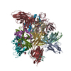
|
|---|---|
| 1 |
|
-Components
-Glycoprotein ... , 2 types, 6 molecules IDMJEN
| #4: Protein | Mass: 31297.158 Da / Num. of mol.: 3 Source method: isolated from a genetically manipulated source Source: (gene. exp.) Ebola virus - Mayinga, Zaire, 1976 / Strain: Mayinga-76 / Gene: GP / Production host:  #5: Protein | Mass: 15335.362 Da / Num. of mol.: 3 Source method: isolated from a genetically manipulated source Source: (gene. exp.) Ebola virus - Mayinga, Zaire, 1976 / Gene: GP, DH33_45401gpGP / Production host: |
|---|
-Antibody , 3 types, 9 molecules FAKGBLHCO
| #1: Antibody | Mass: 13057.619 Da / Num. of mol.: 3 Source method: isolated from a genetically manipulated source Source: (gene. exp.) Homo sapiens (human) / Production host: #2: Antibody | Mass: 11738.831 Da / Num. of mol.: 3 Source method: isolated from a genetically manipulated source Source: (gene. exp.) Homo sapiens (human) / Production host: #3: Antibody | Mass: 25853.459 Da / Num. of mol.: 3 Source method: isolated from a genetically manipulated source Source: (gene. exp.) |
|---|
-Sugars , 2 types, 6 molecules 
| #6: Polysaccharide | Source method: isolated from a genetically manipulated source #7: Sugar |  Type: D-saccharide, beta linking / Mass: 221.208 Da / Num. of mol.: 3 / Source method: obtained synthetically / Formula: C8H15NO6 Type: D-saccharide, beta linking / Mass: 221.208 Da / Num. of mol.: 3 / Source method: obtained synthetically / Formula: C8H15NO6 |
|---|
-Details
| Has ligand of interest | N |
|---|---|
| Has protein modification | Y |
-Experimental details
-Experiment
| Experiment | Method: ELECTRON MICROSCOPY |
|---|---|
| EM experiment | Aggregation state: PARTICLE / 3D reconstruction method: single particle reconstruction |
- Sample preparation
| Component | Name: Structure of EBOV GP in complex with 2.1.1D5 scFv and 6D6 scFv Type: COMPLEX / Entity ID: #1-#5 / Source: RECOMBINANT |
|---|---|
| Source (natural) | Organism: Ebola virus - Mayinga, Zaire, 1976 |
| Source (recombinant) | Organism: |
| Buffer solution | pH: 7.4 / Details: TBS buffer pH 7.4 |
| Specimen | Embedding applied: NO / Shadowing applied: NO / Staining applied: NO / Vitrification applied: YES |
| Vitrification | Cryogen name: ETHANE |
- Electron microscopy imaging
| Experimental equipment |  Model: Titan Krios / Image courtesy: FEI Company |
|---|---|
| Microscopy | Model: FEI TITAN KRIOS |
| Electron gun | Electron source:  FIELD EMISSION GUN / Accelerating voltage: 300 kV / Illumination mode: FLOOD BEAM FIELD EMISSION GUN / Accelerating voltage: 300 kV / Illumination mode: FLOOD BEAM |
| Electron lens | Mode: BRIGHT FIELD / Nominal defocus max: 2500 nm / Nominal defocus min: 1000 nm |
| Image recording | Electron dose: 50 e/Å2 / Film or detector model: GATAN K3 (6k x 4k) |
- Processing
| Software | Name: PHENIX / Version: 1.18.2_3874: / Classification: refinement | ||||||||||||||||||||||||
|---|---|---|---|---|---|---|---|---|---|---|---|---|---|---|---|---|---|---|---|---|---|---|---|---|---|
| EM software | Name: PHENIX / Category: model refinement | ||||||||||||||||||||||||
| CTF correction | Type: PHASE FLIPPING AND AMPLITUDE CORRECTION | ||||||||||||||||||||||||
| 3D reconstruction | Resolution: 2.53 Å / Resolution method: FSC 0.143 CUT-OFF / Num. of particles: 610008 / Symmetry type: POINT | ||||||||||||||||||||||||
| Refine LS restraints |
|
