 Movie
Movie Controller


+ Open data

- Basic information
| Entry | Database: PDB / ID: 8dpd | |||||||||
|---|---|---|---|---|---|---|---|---|---|---|
| Title | superfolder GFP Tyr74pCNPhe mutant | |||||||||
 Components Components | Superfolder green fluorescent protein | |||||||||
 Keywords Keywords | FLUORESCENT PROTEIN / unnatural amino acid / cyanophenylalanine | |||||||||
| Function / homology | Green fluorescent protein, GFP / Green fluorescent protein-related / Green fluorescent protein / Green fluorescent protein / bioluminescence / generation of precursor metabolites and energy / CARBON DIOXIDE / DI(HYDROXYETHYL)ETHER / Green fluorescent protein Function and homology information Function and homology information | |||||||||
| Biological species |   Aequorea victoria (jellyfish) Aequorea victoria (jellyfish) | |||||||||
| Method |  X-RAY DIFFRACTION / SYNCHROTRON / MOLECULAR REPLACEMENT / Resolution: 1.51 Å X-RAY DIFFRACTION / SYNCHROTRON / MOLECULAR REPLACEMENT / Resolution: 1.51 Å | |||||||||
 Authors Authors | Phillips-Piro, C.M. / Papoutsis, B. / Piacentini, J. | |||||||||
| Funding support |  United States, 1items United States, 1items
| |||||||||
 Citation Citation | Journal: To Be Published Title: Superfolder GFP Tyr74pCNPhe mutant Authors: Phillips-Piro, C.M. / Papoutsis, B. / Piacentini, J. | |||||||||
| History |
|
- Structure visualization
| Structure viewer | Molecule: MolmilJmol/JSmol |
|---|
- Downloads & links
-Download
| PDBx/mmCIF format | 8dpd.cif.gz | 134.9 KB | Display | PDBx/mmCIF format |
|---|---|---|---|---|
| PDB format | pdb8dpd.ent.gz | 86 KB | Display | PDB format |
| PDBx/mmJSON format | 8dpd.json.gz | Tree view | PDBx/mmJSON format | |
| Others |  Other downloads Other downloads |
-Validation report
| Arichive directory | https://data.pdbj.org/pub/pdb/validation_reports/dp/8dpdftp://data.pdbj.org/pub/pdb/validation_reports/dp/8dpd | HTTPS FTP |
|---|
-Related structure data
| Related structure data |  2b3pS S: Starting model for refinement |
|---|---|
| Similar structure data |
-Links
 PDBj
PDBj


- Assembly
| Deposited unit | 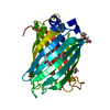
| ||||||||||||
|---|---|---|---|---|---|---|---|---|---|---|---|---|---|
| 1 |
| ||||||||||||
| Unit cell |
|
-Components
-Protein , 1 types, 1 molecules A
| #1: Protein | Mass: 26907.336 Da / Num. of mol.: 1 Mutation: S30R, Y39N, F64L, Q80R, F99S, N105T, Y145F, M153T, V163A, I171V, A206V Source method: isolated from a genetically manipulated source Source: (gene. exp.) Aequorea victoria (jellyfish) / Gene: GFP / Production host:  |
|---|
-Non-polymers , 5 types, 237 molecules 


| #2: Chemical | ChemComp-CO2 /  Mass: 44.010 Da / Num. of mol.: 1 / Source method: obtained synthetically / Formula: CO2 Mass: 44.010 Da / Num. of mol.: 1 / Source method: obtained synthetically / Formula: CO2 | ||||||
|---|---|---|---|---|---|---|---|
| #3: Chemical | ChemComp-EDO /  Mass: 62.068 Da / Num. of mol.: 4 / Source method: obtained synthetically / Formula: C2H6O2 Mass: 62.068 Da / Num. of mol.: 4 / Source method: obtained synthetically / Formula: C2H6O2#4: Chemical | ChemComp-PEG / |  Mass: 106.120 Da / Num. of mol.: 1 / Source method: obtained synthetically / Formula: C4H10O3 Mass: 106.120 Da / Num. of mol.: 1 / Source method: obtained synthetically / Formula: C4H10O3#5: Chemical | ChemComp-MG /  Mass: 24.305 Da / Num. of mol.: 4 / Source method: obtained synthetically / Formula: Mg Mass: 24.305 Da / Num. of mol.: 4 / Source method: obtained synthetically / Formula: Mg#6: Water | ChemComp-HOH / | Mass: 18.015 Da / Num. of mol.: 227 / Source method: isolated from a natural source / Formula: H2O |
-Details
| Has ligand of interest | Y |
|---|---|
| Has protein modification | Y |
-Experimental details
-Experiment
| Experiment | Method: X-RAY DIFFRACTION / Number of used crystals: 1 |
|---|
- Sample preparation
| Crystal | Density Matthews: 2.07 Å3/Da / Density % sol: 40.59 % |
|---|---|
| Crystal grow | Temperature: 298 K / Method: vapor diffusion, sitting drop Details: 0.2 M Magnesium chloride, 0.1 M Tris-HCl pH 8.5, 25% PEG 3350 |
-Data collection
| Diffraction | Mean temperature: 100 K / Serial crystal experiment: N |
|---|---|
| Diffraction source | Source: SYNCHROTRON / Site: APS / Beamline: 24-ID-E / Wavelength: 0.9792 Å |
| Detector | Type: DECTRIS EIGER X 16M / Detector: PIXEL / Date: May 21, 2017 |
| Radiation | Protocol: SINGLE WAVELENGTH / Monochromatic (M) / Laue (L): M / Scattering type: x-ray |
| Radiation wavelength | Wavelength: 0.9792 Å / Relative weight: 1 |
| Reflection | Resolution: 1.51→19.9 Å / Num. obs: 37289 / % possible obs: 99.2 % / Redundancy: 8.3 % / Biso Wilson estimate: 23.78 Å2 / CC1/2: 0.999 / Net I/σ(I): 14.6 |
| Reflection shell | Resolution: 1.51→1.55 Å / Mean I/σ(I) obs: 7.7 / Num. unique obs: 1762 / CC1/2: 0.572 |
- Processing
| Software |
| |||||||||||||||||||||||||||||||||||||||||||||||||||||||||||||||||||||||||||||||||||||||||||||||||||||||||
|---|---|---|---|---|---|---|---|---|---|---|---|---|---|---|---|---|---|---|---|---|---|---|---|---|---|---|---|---|---|---|---|---|---|---|---|---|---|---|---|---|---|---|---|---|---|---|---|---|---|---|---|---|---|---|---|---|---|---|---|---|---|---|---|---|---|---|---|---|---|---|---|---|---|---|---|---|---|---|---|---|---|---|---|---|---|---|---|---|---|---|---|---|---|---|---|---|---|---|---|---|---|---|---|---|---|---|
| Refinement | Method to determine structure: MOLECULAR REPLACEMENT Starting model: 2B3P Resolution: 1.51→19.9 Å / SU ML: 0.2107 / Cross valid method: FREE R-VALUE / σ(F): 1.34 / Phase error: 27.3979 Stereochemistry target values: GeoStd + Monomer Library + CDL v1.2
| |||||||||||||||||||||||||||||||||||||||||||||||||||||||||||||||||||||||||||||||||||||||||||||||||||||||||
| Solvent computation | Shrinkage radii: 0.9 Å / VDW probe radii: 1.11 Å / Solvent model: FLAT BULK SOLVENT MODEL | |||||||||||||||||||||||||||||||||||||||||||||||||||||||||||||||||||||||||||||||||||||||||||||||||||||||||
| Displacement parameters | Biso mean: 35.97 Å2 | |||||||||||||||||||||||||||||||||||||||||||||||||||||||||||||||||||||||||||||||||||||||||||||||||||||||||
| Refinement step | Cycle: LAST / Resolution: 1.51→19.9 Å
| |||||||||||||||||||||||||||||||||||||||||||||||||||||||||||||||||||||||||||||||||||||||||||||||||||||||||
| Refine LS restraints |
| |||||||||||||||||||||||||||||||||||||||||||||||||||||||||||||||||||||||||||||||||||||||||||||||||||||||||
| LS refinement shell |
|
