 Movie
Movie Controller


+ Open data

- Basic information
| Entry | Database: PDB / ID: 8dot | ||||||
|---|---|---|---|---|---|---|---|
| Title | Structure of a methane clathrate binding protein | ||||||
 Components Components | methane clathrate binding protein | ||||||
 Keywords Keywords | ANTIFREEZE PROTEIN / methane / clathrate / subsurface / metagenome | ||||||
| Function / homology | Streptococcal non-M secreted SibA / cytoskeleton / Uncharacterized protein Function and homology information Function and homology information | ||||||
| Biological species | marine sediment metagenome (others) | ||||||
| Method |  X-RAY DIFFRACTION / SYNCHROTRON / MOLECULAR REPLACEMENT / Resolution: 3.05 Å X-RAY DIFFRACTION / SYNCHROTRON / MOLECULAR REPLACEMENT / Resolution: 3.05 Å | ||||||
 Authors Authors | Huard, D.J.E. / Lieberman, R.L. / Glass, J.B. | ||||||
| Funding support |  United States, 1items United States, 1items
| ||||||
 Citation Citation | Journal: Pnas Nexus / Year: 2023 Title: Molecular basis for inhibition of methane clathrate growth by a deep subsurface bacterial protein. Authors: Huard, D.J.E. / Johnson, A.M. / Fan, Z. / Kenney, L.G. / Xu, M. / Drori, R. / Gumbart, J.C. / Dai, S. / Lieberman, R.L. / Glass, J.B. | ||||||
| History |
|
- Structure visualization
| Structure viewer | Molecule: MolmilJmol/JSmol |
|---|
- Downloads & links
-Download
| PDBx/mmCIF format | 8dot.cif.gz | 52.7 KB | Display | PDBx/mmCIF format |
|---|---|---|---|---|
| PDB format | pdb8dot.ent.gz | 32.4 KB | Display | PDB format |
| PDBx/mmJSON format | 8dot.json.gz | Tree view | PDBx/mmJSON format | |
| Others |  Other downloads Other downloads |
-Validation report
| Arichive directory | https://data.pdbj.org/pub/pdb/validation_reports/do/8dotftp://data.pdbj.org/pub/pdb/validation_reports/do/8dot | HTTPS FTP |
|---|
-Related structure data
| Similar structure data |
|---|
-Links
 PDBj
PDBj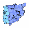
- Assembly
| Deposited unit | 
| ||||||||||||
|---|---|---|---|---|---|---|---|---|---|---|---|---|---|
| 1 |
| ||||||||||||
| Unit cell |
|
-Components
| #1: Protein | Mass: 47183.254 Da / Num. of mol.: 1 Source method: isolated from a genetically manipulated source Source: (gene. exp.) marine sediment metagenome (others) / Gene: S12H4_00347 / Production host:  |
|---|---|
| #2: Chemical | ChemComp-PG4 /   Mass: 194.226 Da / Num. of mol.: 1 / Source method: obtained synthetically / Formula: C8H18O5 / Comment: precipitant*YM Mass: 194.226 Da / Num. of mol.: 1 / Source method: obtained synthetically / Formula: C8H18O5 / Comment: precipitant*YM |
| Has ligand of interest | N |
-Experimental details
-Experiment
| Experiment | Method: X-RAY DIFFRACTION / Number of used crystals: 1 |
|---|
- Sample preparation
| Crystal | Density Matthews: 1.81 Å3/Da / Density % sol: 31.9 % |
|---|---|
| Crystal grow | Temperature: 277 K / Method: vapor diffusion, sitting drop Details: 0.16 M sodium acetate trihydrate, 7.5% PEG 8,000 and 20% glycerol |
-Data collection
| Diffraction | Mean temperature: 100 K / Serial crystal experiment: N |
|---|---|
| Diffraction source | Source: SYNCHROTRON / Site: APS / Beamline: 22-ID / Wavelength: 1 Å |
| Detector | Type: DECTRIS EIGER X 16M / Detector: PIXEL / Date: Jul 28, 2021 |
| Radiation | Protocol: SINGLE WAVELENGTH / Monochromatic (M) / Laue (L): M / Scattering type: x-ray |
| Radiation wavelength | Wavelength: 1 Å / Relative weight: 1 |
| Reflection | Resolution: 3.05→39.05 Å / Num. obs: 7012 / % possible obs: 99.72 % / Redundancy: 25 % / Biso Wilson estimate: 76.1 Å2 / CC1/2: 0.999 / Net I/σ(I): 25.17 |
| Reflection shell | Resolution: 3.05→3.159 Å / Mean I/σ(I) obs: 7.27 / Num. unique obs: 675 / CC1/2: 0.989 |
- Processing
| Software |
| ||||||||||||||||||||||||||||||||||||||||||||||||||||||||||||||||||||||
|---|---|---|---|---|---|---|---|---|---|---|---|---|---|---|---|---|---|---|---|---|---|---|---|---|---|---|---|---|---|---|---|---|---|---|---|---|---|---|---|---|---|---|---|---|---|---|---|---|---|---|---|---|---|---|---|---|---|---|---|---|---|---|---|---|---|---|---|---|---|---|---|
| Refinement | Method to determine structure: MOLECULAR REPLACEMENT Starting model: alphafold Resolution: 3.05→39.05 Å / SU ML: 0.4425 / Cross valid method: FREE R-VALUE / σ(F): 1.38 / Phase error: 24.576 Stereochemistry target values: GeoStd + Monomer Library + CDL v1.2
| ||||||||||||||||||||||||||||||||||||||||||||||||||||||||||||||||||||||
| Solvent computation | Shrinkage radii: 0.9 Å / VDW probe radii: 1.11 Å / Solvent model: FLAT BULK SOLVENT MODEL | ||||||||||||||||||||||||||||||||||||||||||||||||||||||||||||||||||||||
| Displacement parameters | Biso mean: 66.12 Å2 | ||||||||||||||||||||||||||||||||||||||||||||||||||||||||||||||||||||||
| Refinement step | Cycle: LAST / Resolution: 3.05→39.05 Å
| ||||||||||||||||||||||||||||||||||||||||||||||||||||||||||||||||||||||
| Refine LS restraints |
| ||||||||||||||||||||||||||||||||||||||||||||||||||||||||||||||||||||||
| LS refinement shell |
|
