 Movie
Movie Controller

[English] 日本語
 Yorodumi
- PDB-8dor: Crystal structure of Dihydropteridine reductase/oxygen-insensitiv... -
+ Open data

- Basic information
| Entry | Database: PDB / ID: 8dor | |||||||||
|---|---|---|---|---|---|---|---|---|---|---|
| Title | Crystal structure of Dihydropteridine reductase/oxygen-insensitive NAD(P)H nitroreductase from Klebsiella pneumoniae | |||||||||
 Components Components | Dihydropteridine reductase/oxygen-insensitive NAD(P)H nitroreductase | |||||||||
 Keywords Keywords | OXIDOREDUCTASE / SSGCID / STRUCTURAL GENOMICS / SEATTLE STRUCTURAL GENOMICS CENTER FOR INFECTIOUS DISEASE | |||||||||
| Function / homology |  Function and homology information Function and homology information | |||||||||
| Biological species |  Klebsiella pneumoniae (bacteria) Klebsiella pneumoniae (bacteria) | |||||||||
| Method |  X-RAY DIFFRACTION / SYNCHROTRON / MOLECULAR REPLACEMENT / molecular replacement / Resolution: 1.35 Å X-RAY DIFFRACTION / SYNCHROTRON / MOLECULAR REPLACEMENT / molecular replacement / Resolution: 1.35 Å | |||||||||
 Authors Authors | Seattle Structural Genomics Center for Infectious Disease (SSGCID) | |||||||||
| Funding support |  United States, 2items United States, 2items
| |||||||||
 Citation Citation | Journal: Acta Crystallogr.,Sect.F / Year: 2024 Title: Crystal structures of NAD(P)H nitroreductases from Klebsiella pneumoniae. Authors: Kancherla, A.D. / Liu, L. / Tillery, L. / Shek, R. / Craig, J.K. / Machen, A.J. / Seibold, S. / Battaile, K.P. / Fradi, S. / Barrett, L.K. / Subramanian, S. / Myler, P. / Van Voorhis, W.C. / Lovell, S. | |||||||||
| History |
|
- Structure visualization
| Structure viewer | Molecule: MolmilJmol/JSmol |
|---|
- Downloads & links
-Download
| PDBx/mmCIF format | 8dor.cif.gz | 374.4 KB | Display | PDBx/mmCIF format |
|---|---|---|---|---|
| PDB format | pdb8dor.ent.gz | 302 KB | Display | PDB format |
| PDBx/mmJSON format | 8dor.json.gz | Tree view | PDBx/mmJSON format | |
| Others |  Other downloads Other downloads |
-Validation report
| Arichive directory | https://data.pdbj.org/pub/pdb/validation_reports/do/8dorftp://data.pdbj.org/pub/pdb/validation_reports/do/8dor | HTTPS FTP |
|---|
-Related structure data
| Related structure data |  1icrS S: Starting model for refinement |
|---|---|
| Similar structure data |
-Links
 PDBj
PDBj- Assembly
| Deposited unit | 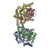
| ||||||||
|---|---|---|---|---|---|---|---|---|---|
| 1 | 
| ||||||||
| 2 | 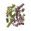
| ||||||||
| Unit cell |
|
-Components
| #1: Protein | Mass: 24959.158 Da / Num. of mol.: 4 / Fragment: KlpnC.20499.a.B1 Source method: isolated from a genetically manipulated source Source: (gene. exp.) Klebsiella pneumoniae (bacteria) / Gene: nfnB, KPN_00553, KPHS_29700 / Production host: #2: Chemical | ChemComp-FMN /   Mass: 456.344 Da / Num. of mol.: 4 / Source method: obtained synthetically / Formula: C17H21N4O9P / Feature type: SUBJECT OF INVESTIGATION Mass: 456.344 Da / Num. of mol.: 4 / Source method: obtained synthetically / Formula: C17H21N4O9P / Feature type: SUBJECT OF INVESTIGATION#3: Chemical | ChemComp-PO4 /   Mass: 94.971 Da / Num. of mol.: 4 / Source method: obtained synthetically / Formula: PO4 Mass: 94.971 Da / Num. of mol.: 4 / Source method: obtained synthetically / Formula: PO4#4: Chemical | ChemComp-GOL /   Mass: 92.094 Da / Num. of mol.: 10 / Source method: obtained synthetically / Formula: C3H8O3 Mass: 92.094 Da / Num. of mol.: 10 / Source method: obtained synthetically / Formula: C3H8O3#5: Water | ChemComp-HOH / |  Mass: 18.015 Da / Num. of mol.: 1077 / Source method: isolated from a natural source / Formula: H2O Mass: 18.015 Da / Num. of mol.: 1077 / Source method: isolated from a natural source / Formula: H2OHas ligand of interest | Y | |
|---|
-Experimental details
-Experiment
| Experiment | Method: X-RAY DIFFRACTION / Number of used crystals: 1 |
|---|
- Sample preparation
| Crystal | Density Matthews: 2.4 Å3/Da / Density % sol: 48.86 % |
|---|---|
| Crystal grow | Temperature: 291 K / Method: vapor diffusion, sitting drop Details: JCSG+ D12: 16% (w/v) PEG 8000; 20% (v/v) glycerol, 0.04 M K2HPO4, KlpnC.20499.a.B1.PW39082 at 30 mg/mL, Tray: plate 12477 well D12 drop 2, Puck: PSL1107, Cryo: DIRECT |
-Data collection
| Diffraction | Mean temperature: 100 K / Serial crystal experiment: N | |||||||||||||||||||||||||||
|---|---|---|---|---|---|---|---|---|---|---|---|---|---|---|---|---|---|---|---|---|---|---|---|---|---|---|---|---|
| Diffraction source | Source: SYNCHROTRON / Site: NSLS-II / Beamline: 19-ID / Wavelength: 0.97949 Å | |||||||||||||||||||||||||||
| Detector | Type: DECTRIS EIGER2 XE 9M / Detector: PIXEL / Date: Mar 24, 2022 | |||||||||||||||||||||||||||
| Radiation | Monochromator: Double Crystal Si 111 / Protocol: SINGLE WAVELENGTH / Monochromatic (M) / Laue (L): M / Scattering type: x-ray | |||||||||||||||||||||||||||
| Radiation wavelength | Wavelength: 0.97949 Å / Relative weight: 1 | |||||||||||||||||||||||||||
| Reflection | Resolution: 1.35→49.21 Å / Num. obs: 204417 / % possible obs: 100 % / Redundancy: 10.8 % / Biso Wilson estimate: 12.3 Å2 / CC1/2: 0.999 / Rmerge(I) obs: 0.101 / Rpim(I) all: 0.032 / Rrim(I) all: 0.106 / Net I/σ(I): 13.5 / Num. measured all: 2214463 / Scaling rejects: 56 | |||||||||||||||||||||||||||
| Reflection shell | Diffraction-ID: 1 / % possible all: 99.9
|
-Phasing
| Phasing | Method: molecular replacement |
|---|
- Processing
| Software |
| ||||||||||||||||||||||||||||||||||||||||||||||||||||||||||||||||||||||||||||||||||||||||||||||||||||||||||||||||||||||||||||||||||||||||||||||||||||||||||||||||||||||||||||||||||||||||||
|---|---|---|---|---|---|---|---|---|---|---|---|---|---|---|---|---|---|---|---|---|---|---|---|---|---|---|---|---|---|---|---|---|---|---|---|---|---|---|---|---|---|---|---|---|---|---|---|---|---|---|---|---|---|---|---|---|---|---|---|---|---|---|---|---|---|---|---|---|---|---|---|---|---|---|---|---|---|---|---|---|---|---|---|---|---|---|---|---|---|---|---|---|---|---|---|---|---|---|---|---|---|---|---|---|---|---|---|---|---|---|---|---|---|---|---|---|---|---|---|---|---|---|---|---|---|---|---|---|---|---|---|---|---|---|---|---|---|---|---|---|---|---|---|---|---|---|---|---|---|---|---|---|---|---|---|---|---|---|---|---|---|---|---|---|---|---|---|---|---|---|---|---|---|---|---|---|---|---|---|---|---|---|---|---|---|---|---|
| Refinement | Method to determine structure: MOLECULAR REPLACEMENT Starting model: 1ICR Resolution: 1.35→29.47 Å / SU ML: 0.11 / Cross valid method: THROUGHOUT / σ(F): 1.96 / Phase error: 16.1 / Stereochemistry target values: ML
| ||||||||||||||||||||||||||||||||||||||||||||||||||||||||||||||||||||||||||||||||||||||||||||||||||||||||||||||||||||||||||||||||||||||||||||||||||||||||||||||||||||||||||||||||||||||||||
| Solvent computation | Shrinkage radii: 0.9 Å / VDW probe radii: 1.11 Å / Solvent model: FLAT BULK SOLVENT MODEL | ||||||||||||||||||||||||||||||||||||||||||||||||||||||||||||||||||||||||||||||||||||||||||||||||||||||||||||||||||||||||||||||||||||||||||||||||||||||||||||||||||||||||||||||||||||||||||
| Displacement parameters | Biso max: 60.34 Å2 / Biso mean: 16.6858 Å2 / Biso min: 6.84 Å2 | ||||||||||||||||||||||||||||||||||||||||||||||||||||||||||||||||||||||||||||||||||||||||||||||||||||||||||||||||||||||||||||||||||||||||||||||||||||||||||||||||||||||||||||||||||||||||||
| Refinement step | Cycle: final / Resolution: 1.35→29.47 Å
| ||||||||||||||||||||||||||||||||||||||||||||||||||||||||||||||||||||||||||||||||||||||||||||||||||||||||||||||||||||||||||||||||||||||||||||||||||||||||||||||||||||||||||||||||||||||||||
| LS refinement shell | Refine-ID: X-RAY DIFFRACTION / Rfactor Rfree error: 0 / Total num. of bins used: 30 / % reflection obs: 100 %
|
