 Movie
Movie Controller

[English] 日本語
 Yorodumi
- PDB-8div: Crystal structure of NavAb I22V as a basis for the human Nav1.7 I... -
+ Open data

- Basic information
| Entry | Database: PDB / ID: 8div | |||||||||||||||
|---|---|---|---|---|---|---|---|---|---|---|---|---|---|---|---|---|
| Title | Crystal structure of NavAb I22V as a basis for the human Nav1.7 Inherited Erythromelalgia I136V mutation | |||||||||||||||
 Components Components | Ion transport protein | |||||||||||||||
 Keywords Keywords | MEMBRANE PROTEIN / Voltage-gated sodium channel Ion transport protein | |||||||||||||||
| Function / homology |  Function and homology information Function and homology informationvoltage-gated sodium channel complex / voltage-gated sodium channel activity / metal ion binding / identical protein binding Similarity search - Function | |||||||||||||||
| Biological species |  Aliarcobacter butzleri RM4018 (bacteria) Aliarcobacter butzleri RM4018 (bacteria) | |||||||||||||||
| Method |  X-RAY DIFFRACTION / SYNCHROTRON / MOLECULAR REPLACEMENT / Resolution: 2.54 Å X-RAY DIFFRACTION / SYNCHROTRON / MOLECULAR REPLACEMENT / Resolution: 2.54 Å | |||||||||||||||
 Authors Authors | Wisedchaisri, G. / Gamal El-Din, T.M. / Powell, N.M. / Zheng, N. / Catterall, W.A. | |||||||||||||||
| Funding support |  United States, 4items United States, 4items
| |||||||||||||||
 Citation Citation | Journal: J.Gen.Physiol. / Year: 2023 Title: Structural basis for severe pain caused by mutations in the voltage sensors of sodium channel NaV1.7. Authors: Wisedchaisri, G. / Gamal El-Din, T.M. / Powell, N.M. / Zheng, N. / Catterall, W.A. | |||||||||||||||
| History |
|
- Structure visualization
| Structure viewer | Molecule: MolmilJmol/JSmol |
|---|
- Downloads & links
-Download
| PDBx/mmCIF format | 8div.cif.gz | 86.6 KB | Display | PDBx/mmCIF format |
|---|---|---|---|---|
| PDB format | pdb8div.ent.gz | 52.3 KB | Display | PDB format |
| PDBx/mmJSON format | 8div.json.gz | Tree view | PDBx/mmJSON format | |
| Others |  Other downloads Other downloads |
-Validation report
| Arichive directory | https://data.pdbj.org/pub/pdb/validation_reports/di/8divftp://data.pdbj.org/pub/pdb/validation_reports/di/8div | HTTPS FTP |
|---|
-Related structure data
| Related structure data |  8diwC  8dixC  8diyC 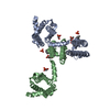 3rvyS S: Starting model for refinement C: citing same article ( |
|---|---|
| Similar structure data |
-Links
 PDBj
PDBj


- Assembly
| Deposited unit | 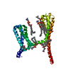
| ||||||||||||
|---|---|---|---|---|---|---|---|---|---|---|---|---|---|
| 1 | 
| ||||||||||||
| Unit cell |
|
-Components
| #1: Protein | Mass: 29775.270 Da / Num. of mol.: 1 / Mutation: I22V Source method: isolated from a genetically manipulated source Source: (gene. exp.) Aliarcobacter butzleri RM4018 (bacteria)Strain: RM4018 / Gene: Abu_1752 / Production host:  Trichoplusia ni (cabbage looper) / References: UniProt: A8EVM5 Trichoplusia ni (cabbage looper) / References: UniProt: A8EVM5 | ||||||||
|---|---|---|---|---|---|---|---|---|---|
| #2: Sugar | ChemComp-BGC /   Type: D-saccharide, beta linking / Mass: 180.156 Da / Num. of mol.: 1 / Source method: obtained synthetically / Formula: C6H12O6 Type: D-saccharide, beta linking / Mass: 180.156 Da / Num. of mol.: 1 / Source method: obtained synthetically / Formula: C6H12O6 | ||||||||
| #3: Chemical | ChemComp-PX4 / 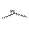  Mass: 678.940 Da / Num. of mol.: 5 / Source method: obtained synthetically / Formula: C36H73NO8P / Comment: DMPC, phospholipid*YM Mass: 678.940 Da / Num. of mol.: 5 / Source method: obtained synthetically / Formula: C36H73NO8P / Comment: DMPC, phospholipid*YM#4: Chemical | 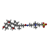  Mass: 614.877 Da / Num. of mol.: 2 / Source method: obtained synthetically / Formula: C32H58N2O7S / Comment: detergent*YM Mass: 614.877 Da / Num. of mol.: 2 / Source method: obtained synthetically / Formula: C32H58N2O7S / Comment: detergent*YM#5: Water | ChemComp-HOH / |  Mass: 18.015 Da / Num. of mol.: 23 / Source method: isolated from a natural source / Formula: H2O Mass: 18.015 Da / Num. of mol.: 23 / Source method: isolated from a natural source / Formula: H2OHas ligand of interest | N | Has protein modification | N | |
-Experimental details
-Experiment
| Experiment | Method: X-RAY DIFFRACTION / Number of used crystals: 1 |
|---|
- Sample preparation
| Crystal | Density Matthews: 6.22 Å3/Da / Density % sol: 80.22 % |
|---|---|
| Crystal grow | Temperature: 277 K / Method: vapor diffusion, hanging drop / pH: 5 Details: 1.7-1.8 M Ammonium sulfate 0.1 M Sodium Citrate pH 5.0 |
-Data collection
| Diffraction | Mean temperature: 100 K / Ambient temp details: Liquid nitrogen / Serial crystal experiment: N |
|---|---|
| Diffraction source | Source: SYNCHROTRON / Site: ALS / Beamline: 8.2.1 / Wavelength: 1 Å |
| Detector | Type: ADSC QUANTUM 315r / Detector: CCD / Date: Mar 7, 2009 |
| Radiation | Monochromator: Double-crystal Si(111) / Protocol: SINGLE WAVELENGTH / Monochromatic (M) / Laue (L): M / Scattering type: x-ray |
| Radiation wavelength | Wavelength: 1 Å / Relative weight: 1 |
| Reflection | Resolution: 2.53→50 Å / Num. obs: 22384 / % possible obs: 87.6 % / Redundancy: 11.5 % / Biso Wilson estimate: 37.29 Å2 / CC1/2: 1 / Rmerge(I) obs: 0.156 / Rpim(I) all: 0.045 / Net I/σ(I): 13.3 |
| Reflection shell | Resolution: 2.53→2.57 Å / Redundancy: 3.6 % / Rmerge(I) obs: 0.844 / Mean I/σ(I) obs: 0.43 / Num. unique obs: 415 / CC1/2: 0.886 / Rpim(I) all: 0.36 / % possible all: 33.3 |
- Processing
| Software |
| ||||||||||||||||||||||||||||||||||||||||||||||||||||||||
|---|---|---|---|---|---|---|---|---|---|---|---|---|---|---|---|---|---|---|---|---|---|---|---|---|---|---|---|---|---|---|---|---|---|---|---|---|---|---|---|---|---|---|---|---|---|---|---|---|---|---|---|---|---|---|---|---|---|
| Refinement | Method to determine structure: MOLECULAR REPLACEMENT Starting model: 3RVY Resolution: 2.54→41.97 Å / SU ML: 0.2531 / Cross valid method: FREE R-VALUE / σ(F): 1.35 / Phase error: 25.4922 Stereochemistry target values: GeoStd + Monomer Library + CDL v1.2
| ||||||||||||||||||||||||||||||||||||||||||||||||||||||||
| Solvent computation | Shrinkage radii: 0.9 Å / VDW probe radii: 1.1 Å / Solvent model: FLAT BULK SOLVENT MODEL | ||||||||||||||||||||||||||||||||||||||||||||||||||||||||
| Displacement parameters | Biso mean: 48.03 Å2 | ||||||||||||||||||||||||||||||||||||||||||||||||||||||||
| Refinement step | Cycle: LAST / Resolution: 2.54→41.97 Å
| ||||||||||||||||||||||||||||||||||||||||||||||||||||||||
| Refine LS restraints |
| ||||||||||||||||||||||||||||||||||||||||||||||||||||||||
| LS refinement shell |
|
