 Movie
Movie Controller


+ Open data

- Basic information
| Entry | Database: PDB / ID: 8dht | ||||||
|---|---|---|---|---|---|---|---|
| Title | Crystal structure of a typeIII Rubisco | ||||||
 Components Components | Ribulose bisphosphate carboxylase | ||||||
 Keywords Keywords | LYASE / Rubisco / 3-phosphoglycerate / complex / PHOTOSYNTHESIS | ||||||
| Function / homology |  Function and homology information Function and homology informationAMP catabolic process / ribulose-bisphosphate carboxylase / ribulose-bisphosphate carboxylase activity / carbon fixation / oxidoreductase activity / magnesium ion binding Similarity search - Function | ||||||
| Biological species |   Archaeoglobus fulgidus (archaea) Archaeoglobus fulgidus (archaea) | ||||||
| Method |  X-RAY DIFFRACTION / SYNCHROTRON / MOLECULAR REPLACEMENT / Resolution: 1.699 Å X-RAY DIFFRACTION / SYNCHROTRON / MOLECULAR REPLACEMENT / Resolution: 1.699 Å | ||||||
 Authors Authors | Qingqiu, H. | ||||||
| Funding support |  United States, 1items United States, 1items
| ||||||
 Citation Citation | Journal: Proteins / Year: 2023 Title: Crystal structure of a type III Rubisco in complex with its product 3-phosphoglycerate. Authors: Huang, Q. / Szebenyi, D.M.E. | ||||||
| History |
|
- Structure visualization
| Structure viewer | Molecule: MolmilJmol/JSmol |
|---|
- Downloads & links
-Download
| PDBx/mmCIF format | 8dht.cif.gz | 379.7 KB | Display | PDBx/mmCIF format |
|---|---|---|---|---|
| PDB format | pdb8dht.ent.gz | 305.7 KB | Display | PDB format |
| PDBx/mmJSON format | 8dht.json.gz | Tree view | PDBx/mmJSON format | |
| Others |  Other downloads Other downloads |
-Validation report
| Arichive directory | https://data.pdbj.org/pub/pdb/validation_reports/dh/8dhtftp://data.pdbj.org/pub/pdb/validation_reports/dh/8dht | HTTPS FTP |
|---|
-Related structure data
| Related structure data |  3kdoS S: Starting model for refinement |
|---|---|
| Similar structure data |
-Links
 PDBj
PDBj

- Assembly
| Deposited unit | 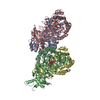
| ||||||||
|---|---|---|---|---|---|---|---|---|---|
| 1 | 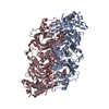
| ||||||||
| 2 | 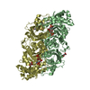
| ||||||||
| Unit cell |
|
-Components
-Protein , 1 types, 4 molecules ABCD
| #1: Protein | Mass: 48695.371 Da / Num. of mol.: 4 Source method: isolated from a genetically manipulated source Source: (gene. exp.) Archaeoglobus fulgidus (archaea) / Gene: rbcL, AF_1638 / Production host:  References: UniProt: O28635, ribulose-bisphosphate carboxylase |
|---|
-Non-polymers , 5 types, 1430 molecules 


| #2: Chemical | ChemComp-MG /  Mass: 24.305 Da / Num. of mol.: 4 / Source method: obtained synthetically / Formula: Mg / Feature type: SUBJECT OF INVESTIGATION Mass: 24.305 Da / Num. of mol.: 4 / Source method: obtained synthetically / Formula: Mg / Feature type: SUBJECT OF INVESTIGATION#3: Chemical | ChemComp-3PG /  Mass: 186.057 Da / Num. of mol.: 8 / Source method: obtained synthetically / Formula: C3H7O7P / Feature type: SUBJECT OF INVESTIGATION Mass: 186.057 Da / Num. of mol.: 8 / Source method: obtained synthetically / Formula: C3H7O7P / Feature type: SUBJECT OF INVESTIGATION#4: Chemical | ChemComp-GOL /  Mass: 92.094 Da / Num. of mol.: 6 / Source method: obtained synthetically / Formula: C3H8O3 / Feature type: SUBJECT OF INVESTIGATION Mass: 92.094 Da / Num. of mol.: 6 / Source method: obtained synthetically / Formula: C3H8O3 / Feature type: SUBJECT OF INVESTIGATION#5: Chemical | ChemComp-ACT /  Mass: 59.044 Da / Num. of mol.: 5 / Source method: obtained synthetically / Formula: C2H3O2 / Feature type: SUBJECT OF INVESTIGATION Mass: 59.044 Da / Num. of mol.: 5 / Source method: obtained synthetically / Formula: C2H3O2 / Feature type: SUBJECT OF INVESTIGATION#6: Water | ChemComp-HOH / | Mass: 18.015 Da / Num. of mol.: 1407 / Source method: isolated from a natural source / Formula: H2O |
|---|
-Details
| Has ligand of interest | Y |
|---|
-Experimental details
-Experiment
| Experiment | Method: X-RAY DIFFRACTION / Number of used crystals: 1 |
|---|
- Sample preparation
| Crystal | Density Matthews: 1.98 Å3/Da / Density % sol: 37.99 % |
|---|---|
| Crystal grow | Temperature: 295 K / Method: vapor diffusion, hanging drop / pH: 8 / Details: 26% PEG4000 (w/v), 0.2M NaAc, 0.1M Tris-HCl, pH8.0 / Temp details: room temperature |
-Data collection
| Diffraction | Mean temperature: 100 K / Serial crystal experiment: N |
|---|---|
| Diffraction source | Source: SYNCHROTRON / Site: CHESS / Beamline: F1 / Wavelength: 0.9775 Å |
| Detector | Type: DECTRIS PILATUS 6M / Detector: PIXEL / Date: May 12, 2018 |
| Radiation | Protocol: SINGLE WAVELENGTH / Monochromatic (M) / Laue (L): M / Scattering type: x-ray |
| Radiation wavelength | Wavelength: 0.9775 Å / Relative weight: 1 |
| Reflection | Resolution: 1.699→50 Å / Num. obs: 166579 / % possible obs: 100 % / Redundancy: 3.4 % / CC1/2: 0.993 / CC star: 0.998 / Rmerge(I) obs: 0.062 / Rpim(I) all: 0.04 / Rrim(I) all: 0.074 / Rsym value: 0.062 / Χ2: 0.619 / Net I/σ(I): 16.5 |
| Reflection shell | Resolution: 1.699→1.73 Å / Redundancy: 3.3 % / Rmerge(I) obs: 0.377 / Mean I/σ(I) obs: 2.087 / Num. unique obs: 8243 / CC1/2: 0.836 / CC star: 0.954 / Rpim(I) all: 0.247 / Rrim(I) all: 0.452 / Rsym value: 0.377 / % possible all: 99.9 |
- Processing
| Software |
| ||||||||||||||||||||||||||||||||||||||||||||||||||||||||||||||||||||||||||||||||||||||||||
|---|---|---|---|---|---|---|---|---|---|---|---|---|---|---|---|---|---|---|---|---|---|---|---|---|---|---|---|---|---|---|---|---|---|---|---|---|---|---|---|---|---|---|---|---|---|---|---|---|---|---|---|---|---|---|---|---|---|---|---|---|---|---|---|---|---|---|---|---|---|---|---|---|---|---|---|---|---|---|---|---|---|---|---|---|---|---|---|---|---|---|---|
| Refinement | Method to determine structure: MOLECULAR REPLACEMENT Starting model: 3KDO Resolution: 1.699→49.377 Å / SU ML: 0.13 / Cross valid method: THROUGHOUT / σ(F): 1.36 / Phase error: 16.1 / Stereochemistry target values: ML
| ||||||||||||||||||||||||||||||||||||||||||||||||||||||||||||||||||||||||||||||||||||||||||
| Solvent computation | Shrinkage radii: 0.9 Å / VDW probe radii: 1.11 Å / Solvent model: FLAT BULK SOLVENT MODEL | ||||||||||||||||||||||||||||||||||||||||||||||||||||||||||||||||||||||||||||||||||||||||||
| Displacement parameters | Biso max: 73.01 Å2 / Biso mean: 21.4751 Å2 / Biso min: 9.36 Å2 | ||||||||||||||||||||||||||||||||||||||||||||||||||||||||||||||||||||||||||||||||||||||||||
| Refinement step | Cycle: final / Resolution: 1.699→49.377 Å
| ||||||||||||||||||||||||||||||||||||||||||||||||||||||||||||||||||||||||||||||||||||||||||
| LS refinement shell | Refine-ID: X-RAY DIFFRACTION / Rfactor Rfree error: 0
|
