 ムービー
ムービー コントローラー


+ データを開く

- 基本情報
基本情報
| 登録情報 | データベース: PDB / ID: 8dhj | ||||||
|---|---|---|---|---|---|---|---|
| タイトル | Crystal structure of Clostridioides difficile Protein Tyrosine Phosphatase at pH 7.5 | ||||||
 要素 要素 | TYR_PHOSPHATASE_2 domain-containing protein | ||||||
 キーワード キーワード | HYDROLASE / phosphatase | ||||||
| 機能・相同性 | Tyrosine/serine-protein phosphatase IphP-type / Tyrosine phosphatase family / phosphoprotein phosphatase activity / Tyrosine specific protein phosphatases domain profile. / Tyrosine-specific protein phosphatases domain / Protein-tyrosine phosphatase-like / Tyrosine specific protein phosphatases domain-containing protein 機能・相同性情報 機能・相同性情報 | ||||||
| 生物種 |  Clostridioides difficile 6050 (バクテリア) Clostridioides difficile 6050 (バクテリア) | ||||||
| 手法 |  X線回折 / シンクロトロン / 分子置換 / 解像度: 1.483 Å X線回折 / シンクロトロン / 分子置換 / 解像度: 1.483 Å | ||||||
 データ登録者 データ登録者 | Lountos, G.T. / Tropea, J.E. | ||||||
| 資金援助 |  米国, 1件 米国, 1件
| ||||||
 引用 引用 | ジャーナル: To Be Published タイトル: Crystal structure of Clostridioides difficile Protein Tyrosine Phosphatase at pH 7.5 著者: Lountos, G.T. / Tropea, J.E. | ||||||
| 履歴 |
|
- 構造の表示
構造の表示
| 構造ビューア | 分子: MolmilJmol/JSmol |
|---|
- ダウンロードとリンク
-ダウンロード
| PDBx/mmCIF形式 | 8dhj.cif.gz | 222 KB | 表示 | PDBx/mmCIF形式 |
|---|---|---|---|---|
| PDB形式 | pdb8dhj.ent.gz | 175.5 KB | 表示 | PDB形式 |
| PDBx/mmJSON形式 | 8dhj.json.gz | ツリー表示 | PDBx/mmJSON形式 | |
| その他 |  その他のダウンロード その他のダウンロード |
-検証レポート
| 文書・要旨 | 8dhj_validation.pdf.gz | 977.1 KB | 表示 | wwPDB検証レポート |
|---|---|---|---|---|
| 文書・詳細版 | 8dhj_full_validation.pdf.gz | 978.9 KB | 表示 | |
| XML形式データ | 8dhj_validation.xml.gz | 24.8 KB | 表示 | |
| CIF形式データ | 8dhj_validation.cif.gz | 37.8 KB | 表示 | |
| アーカイブディレクトリ | https://data.pdbj.org/pub/pdb/validation_reports/dh/8dhjftp://data.pdbj.org/pub/pdb/validation_reports/dh/8dhj | HTTPS FTP |
-関連構造データ
| 関連構造データ |  1ywfS S: 精密化の開始モデル |
|---|---|
| 類似構造データ |
-リンク
 PDBj
PDBj


- 集合体
集合体
| 登録構造単位 | 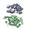
| ||||||||
|---|---|---|---|---|---|---|---|---|---|
| 1 | 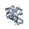
| ||||||||
| 2 | 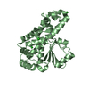
| ||||||||
| 単位格子 |
|
-要素
| #1: タンパク質 | 分子量: 28969.008 Da / 分子数: 2 変異: First three glycine residues are non-native, remnants of linker 由来タイプ: 組換発現 由来: (組換発現) Clostridioides difficile 6050 (バクテリア)プラスミド: pJT510 / 発現宿主: #2: 化合物 |   分子量: 238.305 Da / 分子数: 2 / 由来タイプ: 合成 / 式: C8H18N2O4S / タイプ: SUBJECT OF INVESTIGATION / コメント: pH緩衝剤*YM 分子量: 238.305 Da / 分子数: 2 / 由来タイプ: 合成 / 式: C8H18N2O4S / タイプ: SUBJECT OF INVESTIGATION / コメント: pH緩衝剤*YM#3: 化合物 | ChemComp-CL /   分子量: 35.453 Da / 分子数: 7 / 由来タイプ: 合成 / 式: Cl 分子量: 35.453 Da / 分子数: 7 / 由来タイプ: 合成 / 式: Cl#4: 水 | ChemComp-HOH / |  分子量: 18.015 Da / 分子数: 538 / 由来タイプ: 天然 / 式: H2O 分子量: 18.015 Da / 分子数: 538 / 由来タイプ: 天然 / 式: H2O研究の焦点であるリガンドがあるか | Y | |
|---|
-実験情報
-実験
| 実験 | 手法: X線回折 / 使用した結晶の数: 1 |
|---|
- 試料調製
試料調製
| 結晶 | マシュー密度: 2.19 Å3/Da / 溶媒含有率: 43.83 % |
|---|---|
| 結晶化 | 温度: 293 K / 手法: 蒸気拡散法, ハンギングドロップ法 / pH: 7.5 詳細: 0.1M HEPES pH 7.5, 0.2 magnesium chloride, 25% (w/v) polyethylene glycol 3350 |
-データ収集
| 回折 | 平均測定温度: 100 K / Serial crystal experiment: N |
|---|---|
| 放射光源 | 由来: シンクロトロン / サイト: APS / ビームライン: 22-BM / 波長: 1 Å |
| 検出器 | タイプ: RAYONIX MX300-HS / 検出器: CCD / 日付: 2021年6月18日 |
| 放射 | プロトコル: SINGLE WAVELENGTH / 単色(M)・ラウエ(L): M / 散乱光タイプ: x-ray |
| 放射波長 | 波長: 1 Å / 相対比: 1 |
| 反射 | 解像度: 1.48→50 Å / Num. obs: 82376 / % possible obs: 98.8 % / 冗長度: 2.6 % / CC1/2: 0.996 / Rmerge(I) obs: 0.048 / Rpim(I) all: 0.035 / Net I/σ(I): 19.5 |
| 反射 シェル | 解像度: 1.48→1.5 Å / Rmerge(I) obs: 0.531 / Mean I/σ(I) obs: 2.2 / Num. unique obs: 4129 / CC1/2: 0.679 / Rpim(I) all: 0.41 |
- 解析
解析
| ソフトウェア |
| ||||||||||||||||||||||||||||||||||||||||||||||||||||||||||||||||||||||||||||||||||||||||||||||||||||||||||||||||||||||||||||||||||||||||||||||||||||||||||||||||||||||||||||||
|---|---|---|---|---|---|---|---|---|---|---|---|---|---|---|---|---|---|---|---|---|---|---|---|---|---|---|---|---|---|---|---|---|---|---|---|---|---|---|---|---|---|---|---|---|---|---|---|---|---|---|---|---|---|---|---|---|---|---|---|---|---|---|---|---|---|---|---|---|---|---|---|---|---|---|---|---|---|---|---|---|---|---|---|---|---|---|---|---|---|---|---|---|---|---|---|---|---|---|---|---|---|---|---|---|---|---|---|---|---|---|---|---|---|---|---|---|---|---|---|---|---|---|---|---|---|---|---|---|---|---|---|---|---|---|---|---|---|---|---|---|---|---|---|---|---|---|---|---|---|---|---|---|---|---|---|---|---|---|---|---|---|---|---|---|---|---|---|---|---|---|---|---|---|---|---|
| 精密化 | 構造決定の手法: 分子置換 開始モデル: 1YWF 解像度: 1.483→36.229 Å / SU ML: 0.14 / 交差検証法: THROUGHOUT / σ(F): 1.36 / 位相誤差: 18.67 / 立体化学のターゲット値: ML
| ||||||||||||||||||||||||||||||||||||||||||||||||||||||||||||||||||||||||||||||||||||||||||||||||||||||||||||||||||||||||||||||||||||||||||||||||||||||||||||||||||||||||||||||
| 溶媒の処理 | 減衰半径: 0.9 Å / VDWプローブ半径: 1.11 Å / 溶媒モデル: FLAT BULK SOLVENT MODEL | ||||||||||||||||||||||||||||||||||||||||||||||||||||||||||||||||||||||||||||||||||||||||||||||||||||||||||||||||||||||||||||||||||||||||||||||||||||||||||||||||||||||||||||||
| 原子変位パラメータ | Biso max: 129.47 Å2 / Biso mean: 21.2777 Å2 / Biso min: 5.77 Å2 | ||||||||||||||||||||||||||||||||||||||||||||||||||||||||||||||||||||||||||||||||||||||||||||||||||||||||||||||||||||||||||||||||||||||||||||||||||||||||||||||||||||||||||||||
| 精密化ステップ | サイクル: final / 解像度: 1.483→36.229 Å
| ||||||||||||||||||||||||||||||||||||||||||||||||||||||||||||||||||||||||||||||||||||||||||||||||||||||||||||||||||||||||||||||||||||||||||||||||||||||||||||||||||||||||||||||
| LS精密化 シェル | Refine-ID: X-RAY DIFFRACTION / Rfactor Rfree error: 0
|
