 Movie
Movie Controller


+ Open data

- Basic information
| Entry | Database: PDB / ID: 8dgl | ||||||
|---|---|---|---|---|---|---|---|
| Title | Crystal Structure of the RdfS Excisionase | ||||||
 Components Components | Recombination Directionality Factor RdfS | ||||||
 Keywords Keywords | DNA BINDING PROTEIN / Excisionase / Recombination Directionality Factor / winged helix-turn-helix / superhelix | ||||||
| Function / homology | Helix-turn-helix domain, group 17 / Helix-turn-helix domain / Putative DNA-binding domain superfamily / Helix-turn-helix domain-containing protein Function and homology information Function and homology information | ||||||
| Biological species |  Mesorhizobium japonicum R7A (bacteria) Mesorhizobium japonicum R7A (bacteria) | ||||||
| Method |  X-RAY DIFFRACTION / SYNCHROTRON / MOLECULAR REPLACEMENT / molecular replacement / Resolution: 2.45 Å X-RAY DIFFRACTION / SYNCHROTRON / MOLECULAR REPLACEMENT / molecular replacement / Resolution: 2.45 Å | ||||||
 Authors Authors | Verdonk, C.J. / Ramsay, J.P. / Marshall, A.C. / Bond, C.S. | ||||||
| Funding support |  Australia, 1items Australia, 1items
| ||||||
 Citation Citation | Journal: Acta Crystallogr D Struct Biol / Year: 2022 Title: Crystallographic and X-ray scattering study of RdfS, a recombination directionality factor from an integrative and conjugative element. Authors: Verdonk, C.J. / Marshall, A.C. / Ramsay, J.P. / Bond, C.S. | ||||||
| History |
|
- Structure visualization
| Structure viewer | Molecule: MolmilJmol/JSmol |
|---|
- Downloads & links
-Download
| PDBx/mmCIF format | 8dgl.cif.gz | 72.9 KB | Display | PDBx/mmCIF format |
|---|---|---|---|---|
| PDB format | pdb8dgl.ent.gz | 52.3 KB | Display | PDB format |
| PDBx/mmJSON format | 8dgl.json.gz | Tree view | PDBx/mmJSON format | |
| Others |  Other downloads Other downloads |
-Validation report
| Summary document | 8dgl_validation.pdf.gz | 459.7 KB | Display | wwPDB validaton report |
|---|---|---|---|---|
| Full document | 8dgl_full_validation.pdf.gz | 460.6 KB | Display | |
| Data in XML | 8dgl_validation.xml.gz | 13.5 KB | Display | |
| Data in CIF | 8dgl_validation.cif.gz | 19.3 KB | Display | |
| Arichive directory | https://data.pdbj.org/pub/pdb/validation_reports/dg/8dglftp://data.pdbj.org/pub/pdb/validation_reports/dg/8dgl | HTTPS FTP |
-Related structure data
| Similar structure data | |
|---|---|
| Other databases |
|
-Links
 PDBj
PDBj

- Assembly
| Deposited unit | 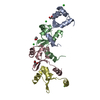
| ||||||||
|---|---|---|---|---|---|---|---|---|---|
| 1 |
| ||||||||
| Unit cell |
|
-Components
| #1: Protein | Mass: 9960.901 Da / Num. of mol.: 4 Source method: isolated from a genetically manipulated source Source: (gene. exp.) Mesorhizobium japonicum R7A (bacteria) / Gene: msi109, A8146_15230, BAE39_30655, EB815_31145 / Plasmid: pETM11 / Production host: #2: Chemical | ChemComp-CL /   Mass: 35.453 Da / Num. of mol.: 4 / Source method: obtained synthetically / Formula: Cl Mass: 35.453 Da / Num. of mol.: 4 / Source method: obtained synthetically / Formula: Cl#3: Chemical |   Mass: 92.094 Da / Num. of mol.: 3 / Source method: obtained synthetically / Formula: C3H8O3 Mass: 92.094 Da / Num. of mol.: 3 / Source method: obtained synthetically / Formula: C3H8O3#4: Water | ChemComp-HOH / |  Mass: 18.015 Da / Num. of mol.: 177 / Source method: isolated from a natural source / Formula: H2O Mass: 18.015 Da / Num. of mol.: 177 / Source method: isolated from a natural source / Formula: H2OHas ligand of interest | N | |
|---|
-Experimental details
-Experiment
| Experiment | Method: X-RAY DIFFRACTION / Number of used crystals: 1 |
|---|
- Sample preparation
| Crystal | Density Matthews: 3.25 Å3/Da / Density meas: 518521.6 Mg/m3 / Density % sol: 62.19 % / Description: Multi-nuclear small needle-like |
|---|---|
| Crystal grow | Temperature: 293 K / Method: vapor diffusion, hanging drop / pH: 6.5 Details: 0.05 M MES; pH 6.5, 4% w/v PEG 5000 MME, 5% v/v 1-propanol, 0.1 M sodium citrate; RdfS protein at 4.3 mg/mL |
-Data collection
| Diffraction | Mean temperature: 100 K / Serial crystal experiment: N | ||||||||||||||||||||||||||||||
|---|---|---|---|---|---|---|---|---|---|---|---|---|---|---|---|---|---|---|---|---|---|---|---|---|---|---|---|---|---|---|---|
| Diffraction source | Source: SYNCHROTRON / Site: Australian Synchrotron / Beamline: MX2 / Wavelength: 0.953 Å | ||||||||||||||||||||||||||||||
| Detector | Type: DECTRIS EIGER X 16M / Detector: PIXEL / Date: Oct 26, 2021 | ||||||||||||||||||||||||||||||
| Radiation | Protocol: SINGLE WAVELENGTH / Monochromatic (M) / Laue (L): M / Scattering type: x-ray | ||||||||||||||||||||||||||||||
| Radiation wavelength | Wavelength: 0.953 Å / Relative weight: 1 | ||||||||||||||||||||||||||||||
| Reflection | Resolution: 2.45→42.87 Å / Num. obs: 19949 / % possible obs: 99.4 % / Redundancy: 6.6 % / Biso Wilson estimate: 41.59 Å2 / CC1/2: 0.993 / Rmerge(I) obs: 0.142 / Rpim(I) all: 0.06 / Rrim(I) all: 0.155 / Net I/σ(I): 9 / Num. measured all: 132047 / Scaling rejects: 21 | ||||||||||||||||||||||||||||||
| Reflection shell | Diffraction-ID: 1
|
-Phasing
| Phasing | Method: molecular replacement | |||||||||
|---|---|---|---|---|---|---|---|---|---|---|
| Phasing MR | Model details: Phaser MODE: MR_AUTO
|
- Processing
| Software |
| ||||||||||||||||||||||||||||||||||||||||||||||||||||||||
|---|---|---|---|---|---|---|---|---|---|---|---|---|---|---|---|---|---|---|---|---|---|---|---|---|---|---|---|---|---|---|---|---|---|---|---|---|---|---|---|---|---|---|---|---|---|---|---|---|---|---|---|---|---|---|---|---|---|
| Refinement | Method to determine structure: MOLECULAR REPLACEMENT Starting model: alphafold prediction Resolution: 2.45→42.87 Å / SU ML: 0.39 / Cross valid method: THROUGHOUT / σ(F): 1.34 / Phase error: 24.78 / Stereochemistry target values: ML
| ||||||||||||||||||||||||||||||||||||||||||||||||||||||||
| Solvent computation | Shrinkage radii: 0.9 Å / VDW probe radii: 1.11 Å / Solvent model: FLAT BULK SOLVENT MODEL | ||||||||||||||||||||||||||||||||||||||||||||||||||||||||
| Displacement parameters | Biso max: 97.6 Å2 / Biso mean: 44.7947 Å2 / Biso min: 23.63 Å2 | ||||||||||||||||||||||||||||||||||||||||||||||||||||||||
| Refinement step | Cycle: final / Resolution: 2.45→42.87 Å
| ||||||||||||||||||||||||||||||||||||||||||||||||||||||||
| LS refinement shell | Refine-ID: X-RAY DIFFRACTION / Rfactor Rfree error: 0 / Total num. of bins used: 7
|
