 ムービー
ムービー コントローラー


+ データを開く

- 基本情報
基本情報
| 登録情報 | データベース: PDB / ID: 8dg4 | ||||||
|---|---|---|---|---|---|---|---|
| タイトル | Group A streptococcus Enolase K252A, K255A, K434A, K435A mutant | ||||||
 要素 要素 | Enolase | ||||||
 キーワード キーワード | LYASE / metalloenzyme / hPg-receptor | ||||||
| 機能・相同性 |  機能・相同性情報 機能・相同性情報phosphopyruvate hydratase / phosphopyruvate hydratase complex / phosphopyruvate hydratase activity / peptidoglycan-based cell wall / glycolytic process / magnesium ion binding / cell surface / extracellular region 類似検索 - 分子機能 | ||||||
| 生物種 |  Streptococcus sp. 'group A' (バクテリア) Streptococcus sp. 'group A' (バクテリア) | ||||||
| 手法 | 電子顕微鏡法 / 単粒子再構成法 / クライオ電子顕微鏡法 / 解像度: 3.12 Å | ||||||
 データ登録者 データ登録者 | Tjia-Fleck, S.C. / Readnour, B.M. / Castellino, F.J. | ||||||
| 資金援助 |  米国, 1件 米国, 1件
| ||||||
 引用 引用 | ジャーナル: Biochemistry / 年: 2023 タイトル: High-Resolution Single-Particle Cryo-EM Hydrated Structure of Enolase Offers Insights into Its Function as a Plasminogen Receptor. 著者: Sheiny Tjia-Fleck / Bradley M Readnour / Yetunde A Ayinuola / Francis J Castellino / 要旨: Cellular plasminogen (Pg) receptors (PgRs) are utilized to recruit Pg; stimulate its activation to the serine protease, plasmin (Pm); and sterically protect the surface Pm from inactivation by host ...Cellular plasminogen (Pg) receptors (PgRs) are utilized to recruit Pg; stimulate its activation to the serine protease, plasmin (Pm); and sterically protect the surface Pm from inactivation by host inhibitors. One such PgR is the moonlighting enzyme, enolase, some of which leaves the cytoplasm and resides at the cell surface to potentially function as a PgR. Since microbes employ conscription of host Pg by PgRs as one virulence mechanism, we explored the structural basis of the ability of enolase (Sen) to function in this manner. Employing single-particle cryo-electron microscopy (cryo-EM), recombinant Sen from was modeled at 2.6 Å as a stable symmetrical doughnut-shaped homooctamer with point group 422 (D4) symmetry, with a monomeric subunit molecular weight of ∼49 kDa. Binding sites for hPg were reported in other studies to include an internal K and the COOH-terminal K residues of Sen. However, in native Sen, the latter are buried within the minor interfaces of the octamer and do not function as a Pg-binding epitope. Whereas Sen and hPg do not interact in solution, when Sen is bound to a surface, hPg interacts with Sen independently of K. PgRs devoid of COOH-terminal lysine utilize lysine isosteres comprising a basic residue, "", and an anionic residue at " + 3" around one turn of an α-helix. We highlight a number of surface-exposed potential hPg-binding lysine isosteres and further conclude that while the octameric structure of Sen is critical for hPg binding, disruption of this octamer without dissociation exposes hPg-binding epitopes. | ||||||
| 履歴 |
|
- 構造の表示
構造の表示
| 構造ビューア | 分子: MolmilJmol/JSmol |
|---|
- ダウンロードとリンク
-ダウンロード
| PDBx/mmCIF形式 | 8dg4.cif.gz | 1 MB | 表示 | PDBx/mmCIF形式 |
|---|---|---|---|---|
| PDB形式 | pdb8dg4.ent.gz | 893.1 KB | 表示 | PDB形式 |
| PDBx/mmJSON形式 | 8dg4.json.gz | ツリー表示 | PDBx/mmJSON形式 | |
| その他 |  その他のダウンロード その他のダウンロード |
-検証レポート
| 文書・要旨 | 8dg4_validation.pdf.gz | 1.3 MB | 表示 | wwPDB検証レポート |
|---|---|---|---|---|
| 文書・詳細版 | 8dg4_full_validation.pdf.gz | 1.3 MB | 表示 | |
| XML形式データ | 8dg4_validation.xml.gz | 89.9 KB | 表示 | |
| CIF形式データ | 8dg4_validation.cif.gz | 141.5 KB | 表示 | |
| アーカイブディレクトリ | https://data.pdbj.org/pub/pdb/validation_reports/dg/8dg4ftp://data.pdbj.org/pub/pdb/validation_reports/dg/8dg4 | HTTPS FTP |
-関連構造データ


-リンク
 PDBj
PDBj
- 集合体
集合体
| 登録構造単位 | 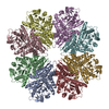
|
|---|---|
| 1 |
|
-要素
| #1: タンパク質 | 分子量: 47310.879 Da / 分子数: 8 / 変異: K252A, K255A, K434A, K435A / 由来タイプ: 組換発現 由来: (組換発現) Streptococcus sp. 'group A' (バクテリア)遺伝子: eno, SPy_0731, M5005_Spy0556 / 発現宿主: |
|---|
-実験情報
-実験
| 実験 | 手法: 電子顕微鏡法 |
|---|---|
| EM実験 | 試料の集合状態: PARTICLE / 3次元再構成法: 単粒子再構成法 |
- 試料調製
試料調製
| 構成要素 | 名称: Octameric Structure of Enolase from Streptococcus Pyogenes タイプ: COMPLEX / Entity ID: all / 由来: RECOMBINANT |
|---|---|
| 分子量 | 値: .396 MDa / 実験値: YES |
| 由来(天然) | 生物種: Streptococcus pyogenes (化膿レンサ球菌) / 株: AP53 |
| 由来(組換発現) | 生物種: |
| 緩衝液 | pH: 7.4 |
| 緩衝液成分 | 濃度: 0.050 mM / 名称: Sodium Phospate / 式: NaH2PO4 |
| 試料 | 包埋: NO / シャドウイング: NO / 染色: NO / 凍結: YES |
| 試料支持 | グリッドのタイプ: Quantifoil R1.2/1.3 |
| 急速凍結 | 装置: FEI VITROBOT MARK IV / 凍結剤: ETHANE / 湿度: 100 % / 凍結前の試料温度: 277 K |
- 電子顕微鏡撮影
| 実験機器 |  モデル: Titan Krios / 画像提供: FEI Company |
|---|---|
| 顕微鏡 | モデル: TFS KRIOS |
| 電子銃 | 電子線源:  FIELD EMISSION GUN / 加速電圧: 300 kV / 照射モード: SPOT SCAN FIELD EMISSION GUN / 加速電圧: 300 kV / 照射モード: SPOT SCAN |
| 電子レンズ | モード: BRIGHT FIELD / 最大 デフォーカス(公称値): 3200 nm / 最小 デフォーカス(公称値): 1100 nm / Cs: 2.7 mm |
| 試料ホルダ | 凍結剤: NITROGEN |
| 撮影 | 電子線照射量: 61.37 e/Å2 フィルム・検出器のモデル: GATAN K2 QUANTUM (4k x 4k) 撮影したグリッド数: 1 / 実像数: 2756 |
| 電子光学装置 | 位相板: VOLTA PHASE PLATE |
- 解析
解析
| EMソフトウェア |
| ||||||||||||||||||||||||
|---|---|---|---|---|---|---|---|---|---|---|---|---|---|---|---|---|---|---|---|---|---|---|---|---|---|
| CTF補正 | タイプ: PHASE FLIPPING AND AMPLITUDE CORRECTION | ||||||||||||||||||||||||
| 粒子像の選択 | 選択した粒子像数: 20000000 | ||||||||||||||||||||||||
| 対称性 | 点対称性: D4 (2回x4回 2面回転対称) | ||||||||||||||||||||||||
| 3次元再構成 | 解像度: 3.12 Å / 解像度の算出法: FSC 0.33 CUT-OFF / 粒子像の数: 813556 / 対称性のタイプ: POINT |
