 Movie
Movie Controller


+ Open data

- Basic information
| Entry | Database: PDB / ID: 8dg4 | ||||||
|---|---|---|---|---|---|---|---|
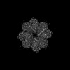
| Title | Group A streptococcus Enolase K252A, K255A, K434A, K435A mutant | ||||||
 Components Components | Enolase | ||||||
 Keywords Keywords | LYASE / metalloenzyme / hPg-receptor | ||||||
| Function / homology |  Function and homology information Function and homology informationphosphopyruvate hydratase / phosphopyruvate hydratase complex / phosphopyruvate hydratase activity / peptidoglycan-based cell wall / glycolytic process / magnesium ion binding / cell surface / extracellular region Similarity search - Function | ||||||
| Biological species |  Streptococcus sp. 'group A' (bacteria) Streptococcus sp. 'group A' (bacteria) | ||||||
| Method | ELECTRON MICROSCOPY / single particle reconstruction / cryo EM / Resolution: 3.12 Å | ||||||
 Authors Authors | Tjia-Fleck, S.C. / Readnour, B.M. / Castellino, F.J. | ||||||
| Funding support |  United States, 1items United States, 1items
| ||||||
 Citation Citation | Journal: Biochemistry / Year: 2023 Title: High-Resolution Single-Particle Cryo-EM Hydrated Structure of Enolase Offers Insights into Its Function as a Plasminogen Receptor. Authors: Sheiny Tjia-Fleck / Bradley M Readnour / Yetunde A Ayinuola / Francis J Castellino / Abstract: Cellular plasminogen (Pg) receptors (PgRs) are utilized to recruit Pg; stimulate its activation to the serine protease, plasmin (Pm); and sterically protect the surface Pm from inactivation by host ...Cellular plasminogen (Pg) receptors (PgRs) are utilized to recruit Pg; stimulate its activation to the serine protease, plasmin (Pm); and sterically protect the surface Pm from inactivation by host inhibitors. One such PgR is the moonlighting enzyme, enolase, some of which leaves the cytoplasm and resides at the cell surface to potentially function as a PgR. Since microbes employ conscription of host Pg by PgRs as one virulence mechanism, we explored the structural basis of the ability of enolase (Sen) to function in this manner. Employing single-particle cryo-electron microscopy (cryo-EM), recombinant Sen from was modeled at 2.6 Å as a stable symmetrical doughnut-shaped homooctamer with point group 422 (D4) symmetry, with a monomeric subunit molecular weight of ∼49 kDa. Binding sites for hPg were reported in other studies to include an internal K and the COOH-terminal K residues of Sen. However, in native Sen, the latter are buried within the minor interfaces of the octamer and do not function as a Pg-binding epitope. Whereas Sen and hPg do not interact in solution, when Sen is bound to a surface, hPg interacts with Sen independently of K. PgRs devoid of COOH-terminal lysine utilize lysine isosteres comprising a basic residue, "", and an anionic residue at " + 3" around one turn of an α-helix. We highlight a number of surface-exposed potential hPg-binding lysine isosteres and further conclude that while the octameric structure of Sen is critical for hPg binding, disruption of this octamer without dissociation exposes hPg-binding epitopes. | ||||||
| History |
|
- Structure visualization
| Structure viewer | Molecule: MolmilJmol/JSmol |
|---|
- Downloads & links
-Download
| PDBx/mmCIF format | 8dg4.cif.gz | 1 MB | Display | PDBx/mmCIF format |
|---|---|---|---|---|
| PDB format | pdb8dg4.ent.gz | 893.1 KB | Display | PDB format |
| PDBx/mmJSON format | 8dg4.json.gz | Tree view | PDBx/mmJSON format | |
| Others |  Other downloads Other downloads |
-Validation report
| Arichive directory | https://data.pdbj.org/pub/pdb/validation_reports/dg/8dg4ftp://data.pdbj.org/pub/pdb/validation_reports/dg/8dg4 | HTTPS FTP |
|---|
-Related structure data
| Related structure data |  27407MC  7uguC C: citing same article ( M: map data used to model this data |
|---|---|
| Similar structure data |
-Links
 PDBj
PDBj
- Assembly
| Deposited unit | 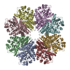
|
|---|---|
| 1 |
|
-Components
| #1: Protein | Mass: 47310.879 Da / Num. of mol.: 8 / Mutation: K252A, K255A, K434A, K435A Source method: isolated from a genetically manipulated source Source: (gene. exp.) Streptococcus sp. 'group A' (bacteria) / Gene: eno, SPy_0731, M5005_Spy0556 / Production host: |
|---|
-Experimental details
-Experiment
| Experiment | Method: ELECTRON MICROSCOPY |
|---|---|
| EM experiment | Aggregation state: PARTICLE / 3D reconstruction method: single particle reconstruction |
- Sample preparation
| Component | Name: Octameric Structure of Enolase from Streptococcus Pyogenes Type: COMPLEX / Entity ID: all / Source: RECOMBINANT |
|---|---|
| Molecular weight | Value: .396 MDa / Experimental value: YES |
| Source (natural) | Organism: Streptococcus pyogenes (bacteria) / Strain: AP53 |
| Source (recombinant) | Organism: |
| Buffer solution | pH: 7.4 |
| Buffer component | Conc.: 0.050 mM / Name: Sodium Phospate / Formula: NaH2PO4 |
| Specimen | Embedding applied: NO / Shadowing applied: NO / Staining applied: NO / Vitrification applied: YES |
| Specimen support | Grid type: Quantifoil R1.2/1.3 |
| Vitrification | Instrument: FEI VITROBOT MARK IV / Cryogen name: ETHANE / Humidity: 100 % / Chamber temperature: 277 K |
- Electron microscopy imaging
| Experimental equipment |  Model: Titan Krios / Image courtesy: FEI Company |
|---|---|
| Microscopy | Model: TFS KRIOS |
| Electron gun | Electron source:  FIELD EMISSION GUN / Accelerating voltage: 300 kV / Illumination mode: SPOT SCAN FIELD EMISSION GUN / Accelerating voltage: 300 kV / Illumination mode: SPOT SCAN |
| Electron lens | Mode: BRIGHT FIELD / Nominal defocus max: 3200 nm / Nominal defocus min: 1100 nm / Cs: 2.7 mm |
| Specimen holder | Cryogen: NITROGEN |
| Image recording | Electron dose: 61.37 e/Å2 / Film or detector model: GATAN K2 QUANTUM (4k x 4k) / Num. of grids imaged: 1 / Num. of real images: 2756 |
| EM imaging optics | Phase plate: VOLTA PHASE PLATE |
- Processing
| EM software |
| ||||||||||||||||||||||||
|---|---|---|---|---|---|---|---|---|---|---|---|---|---|---|---|---|---|---|---|---|---|---|---|---|---|
| CTF correction | Type: PHASE FLIPPING AND AMPLITUDE CORRECTION | ||||||||||||||||||||||||
| Particle selection | Num. of particles selected: 20000000 | ||||||||||||||||||||||||
| Symmetry | Point symmetry: D4 (2x4 fold dihedral) | ||||||||||||||||||||||||
| 3D reconstruction | Resolution: 3.12 Å / Resolution method: FSC 0.33 CUT-OFF / Num. of particles: 813556 / Symmetry type: POINT |
