 Movie
Movie Controller


+ Open data

- Basic information
| Entry | Database: PDB / ID: 8db9 | ||||||
|---|---|---|---|---|---|---|---|
| Title | Adenosine/guanosine nucleoside hydrolase bound to inhibitor | ||||||
 Components Components | Inosine-uridine preferring nucleoside hydrolase family protein | ||||||
 Keywords Keywords | HYDROLASE/HYDROLASE inhibitor / Nucleoside / Hydrolase / Adenosine / Guanosine / Parasitic / Inhibitor / HYDROLASE-HYDROLASE inhibitor complex | ||||||
| Function / homology |  Function and homology information Function and homology informationpurine nucleosidase activity / purine nucleoside catabolic process / metal ion binding / cytosol Similarity search - Function | ||||||
| Biological species |  Trichomonas vaginalis (eukaryote) Trichomonas vaginalis (eukaryote) | ||||||
| Method |  X-RAY DIFFRACTION / SYNCHROTRON / MOLECULAR REPLACEMENT / Resolution: 2.89 Å X-RAY DIFFRACTION / SYNCHROTRON / MOLECULAR REPLACEMENT / Resolution: 2.89 Å | ||||||
 Authors Authors | Muellers, S.N. / Allen, K.N. / Stockman, B.J. | ||||||
| Funding support |  United States, 1items United States, 1items
| ||||||
 Citation Citation | Journal: Biochemistry / Year: 2022 Title: Structure-Guided Insight into the Specificity and Mechanism of a Parasitic Nucleoside Hydrolase. Authors: Muellers, S.N. / Nyitray, M.M. / Reynarowych, N. / Saljanin, E. / Benzie, A.L. / Schoenfeld, A.R. / Stockman, B.J. / Allen, K.N. | ||||||
| History |
|
- Structure visualization
| Structure viewer | Molecule: MolmilJmol/JSmol |
|---|
- Downloads & links
-Download
| PDBx/mmCIF format | 8db9.cif.gz | 238.3 KB | Display | PDBx/mmCIF format |
|---|---|---|---|---|
| PDB format | pdb8db9.ent.gz | 189.2 KB | Display | PDB format |
| PDBx/mmJSON format | 8db9.json.gz | Tree view | PDBx/mmJSON format | |
| Others |  Other downloads Other downloads |
-Validation report
| Arichive directory | https://data.pdbj.org/pub/pdb/validation_reports/db/8db9ftp://data.pdbj.org/pub/pdb/validation_reports/db/8db9 | HTTPS FTP |
|---|
-Related structure data
-Links
 PDBj
PDBj- Assembly
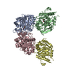
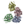
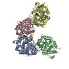
| Deposited unit | 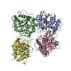
| ||||||||||||
|---|---|---|---|---|---|---|---|---|---|---|---|---|---|
| 1 | 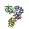
| ||||||||||||
| Unit cell |
|
-Components
| #1: Protein | Mass: 33476.336 Da / Num. of mol.: 4 Source method: isolated from a genetically manipulated source Source: (gene. exp.) Trichomonas vaginalis (eukaryote) / Gene: TVAG_213720 / Production host:  #2: Chemical | ChemComp-CA /   Mass: 40.078 Da / Num. of mol.: 4 / Source method: obtained synthetically / Formula: Ca Mass: 40.078 Da / Num. of mol.: 4 / Source method: obtained synthetically / Formula: Ca#3: Chemical | 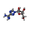  Mass: 243.220 Da / Num. of mol.: 3 / Source method: obtained synthetically / Formula: C8H13N5O4 / Feature type: SUBJECT OF INVESTIGATION Mass: 243.220 Da / Num. of mol.: 3 / Source method: obtained synthetically / Formula: C8H13N5O4 / Feature type: SUBJECT OF INVESTIGATIONHas ligand of interest | Y | |
|---|
-Experimental details
-Experiment
| Experiment | Method: X-RAY DIFFRACTION / Number of used crystals: 1 |
|---|
- Sample preparation
| Crystal | Density Matthews: 2.25 Å3/Da / Density % sol: 45.26 % |
|---|---|
| Crystal grow | Temperature: 290 K / Method: vapor diffusion, hanging drop Details: 0.2 M ammonium sulfate, 25% PEG-3350, and 0.1 M Bis-tris, pH 5.5 |
-Data collection
| Diffraction | Mean temperature: 100 K / Serial crystal experiment: N |
|---|---|
| Diffraction source | Source: SYNCHROTRON / Site: SSRL / Beamline: BL12-2 / Wavelength: 0.977 Å |
| Detector | Type: DECTRIS PILATUS 6M / Detector: PIXEL / Date: Feb 25, 2022 |
| Radiation | Protocol: SINGLE WAVELENGTH / Monochromatic (M) / Laue (L): M / Scattering type: x-ray |
| Radiation wavelength | Wavelength: 0.977 Å / Relative weight: 1 |
| Reflection | Resolution: 2.89→38.13 Å / Num. obs: 23552 / % possible obs: 87.89 % / Redundancy: 4.2 % / Biso Wilson estimate: 61.28 Å2 / CC1/2: 0.988 / Rmerge(I) obs: 0.192 / Rpim(I) all: 0.105 / Rrim(I) all: 0.22 / Net I/σ(I): 6.9 |
| Reflection shell | Resolution: 2.89→2.993 Å / Redundancy: 3.7 % / Rmerge(I) obs: 0.129 / Mean I/σ(I) obs: 1.1 / Num. unique obs: 2425 / CC1/2: 0.408 / Rpim(I) all: 0.738 / Rrim(I) all: 0.149 / % possible all: 88.09 |
- Processing
| Software |
| |||||||||||||||||||||||||||||||||||||||||||||||||||||||||||||||||||||||||||||
|---|---|---|---|---|---|---|---|---|---|---|---|---|---|---|---|---|---|---|---|---|---|---|---|---|---|---|---|---|---|---|---|---|---|---|---|---|---|---|---|---|---|---|---|---|---|---|---|---|---|---|---|---|---|---|---|---|---|---|---|---|---|---|---|---|---|---|---|---|---|---|---|---|---|---|---|---|---|---|
| Refinement | Method to determine structure: MOLECULAR REPLACEMENT Starting model: AlphaFold model Resolution: 2.89→38.13 Å / SU ML: 0.587 / Cross valid method: FREE R-VALUE / σ(F): 1.96 / Phase error: 39.3732 Stereochemistry target values: GeoStd + Monomer Library + CDL v1.2
| |||||||||||||||||||||||||||||||||||||||||||||||||||||||||||||||||||||||||||||
| Solvent computation | Shrinkage radii: 0.9 Å / VDW probe radii: 1.11 Å / Solvent model: FLAT BULK SOLVENT MODEL | |||||||||||||||||||||||||||||||||||||||||||||||||||||||||||||||||||||||||||||
| Displacement parameters | Biso mean: 54.8 Å2 | |||||||||||||||||||||||||||||||||||||||||||||||||||||||||||||||||||||||||||||
| Refinement step | Cycle: LAST / Resolution: 2.89→38.13 Å
| |||||||||||||||||||||||||||||||||||||||||||||||||||||||||||||||||||||||||||||
| Refine LS restraints |
| |||||||||||||||||||||||||||||||||||||||||||||||||||||||||||||||||||||||||||||
| LS refinement shell |
|
