 Movie
Movie Controller


+ Open data

- Basic information
| Entry | Database: PDB / ID: 8da5 | |||||||||
|---|---|---|---|---|---|---|---|---|---|---|
| Title | Coevolved affibody-Z domain pair LL1.c4 | |||||||||
 Components Components |
| |||||||||
 Keywords Keywords | PROTEIN BINDING / affibody | |||||||||
| Function / homology |  Function and homology information Function and homology information | |||||||||
| Biological species |   Staphylococcus aureus (bacteria) Staphylococcus aureus (bacteria)synthetic construct (others) | |||||||||
| Method |  X-RAY DIFFRACTION / SYNCHROTRON / MOLECULAR REPLACEMENT / Resolution: 1 Å X-RAY DIFFRACTION / SYNCHROTRON / MOLECULAR REPLACEMENT / Resolution: 1 Å | |||||||||
 Authors Authors | Jude, K.M. / Yang, A. / Garcia, K.C. | |||||||||
| Funding support |  United States, 2items United States, 2items
| |||||||||
 Citation Citation | Journal: Science / Year: 2023 Title: Deploying synthetic coevolution and machine learning to engineer protein-protein interactions. Authors: Yang, A. / Jude, K.M. / Lai, B. / Minot, M. / Kocyla, A.M. / Glassman, C.R. / Nishimiya, D. / Kim, Y.S. / Reddy, S.T. / Khan, A.A. / Garcia, K.C. | |||||||||
| History |
|
- Structure visualization
| Structure viewer | Molecule: MolmilJmol/JSmol |
|---|
- Downloads & links
-Download
| PDBx/mmCIF format | 8da5.cif.gz | 193 KB | Display | PDBx/mmCIF format |
|---|---|---|---|---|
| PDB format | pdb8da5.ent.gz | 132.4 KB | Display | PDB format |
| PDBx/mmJSON format | 8da5.json.gz | Tree view | PDBx/mmJSON format | |
| Others |  Other downloads Other downloads |
-Validation report
| Arichive directory | https://data.pdbj.org/pub/pdb/validation_reports/da/8da5ftp://data.pdbj.org/pub/pdb/validation_reports/da/8da5 | HTTPS FTP |
|---|
-Related structure data
| Related structure data |  8da3C  8da4C  8da6C  8da7C  8da8C  8da9C  8daaC  8dabC  8dacC 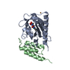 5djtS C: citing same article ( S: Starting model for refinement |
|---|---|
| Similar structure data | |
| Experimental dataset #1 | Data reference: 10.15785/SBGRID/922 / Data set type: diffraction image data |
-Links
 PDBj
PDBj


- Assembly
| Deposited unit | 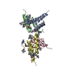
| ||||||||||||
|---|---|---|---|---|---|---|---|---|---|---|---|---|---|
| 1 | 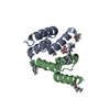
| ||||||||||||
| 2 | 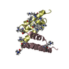
| ||||||||||||
| Unit cell |
|
-Components
-Affibody LL1. ... , 2 types, 2 molecules BD
| #3: Protein | Mass: 7715.715 Da / Num. of mol.: 1 Source method: isolated from a genetically manipulated source Source: (gene. exp.) synthetic construct (others) / Production host: |
|---|---|
| #4: Protein | Mass: 7743.769 Da / Num. of mol.: 1 Source method: isolated from a genetically manipulated source Source: (gene. exp.) synthetic construct (others) / Production host: |
-Antibody , 2 types, 2 molecules AC
| #1: Antibody | Mass: 7864.690 Da / Num. of mol.: 1 / Mutation: Q9F, F13I, G29A, I31F Source method: isolated from a genetically manipulated source Source: (gene. exp.) Staphylococcus aureus (bacteria) / Gene: spa / Production host: |
|---|---|
| #2: Antibody | Mass: 7918.783 Da / Num. of mol.: 1 / Mutation: Q9F, F13I, G29A, I31F Source method: isolated from a genetically manipulated source Source: (gene. exp.) Staphylococcus aureus (bacteria) / Gene: spa / Production host: |
-Non-polymers , 2 types, 380 molecules 

| #5: Chemical | ChemComp-GOL /  Mass: 92.094 Da / Num. of mol.: 1 / Source method: obtained synthetically / Formula: C3H8O3 Mass: 92.094 Da / Num. of mol.: 1 / Source method: obtained synthetically / Formula: C3H8O3 |
|---|---|
| #6: Water | ChemComp-HOH / Mass: 18.015 Da / Num. of mol.: 379 / Source method: isolated from a natural source / Formula: H2O |
-Details
| Has ligand of interest | N |
|---|
-Experimental details
-Experiment
| Experiment | Method: X-RAY DIFFRACTION / Number of used crystals: 1 |
|---|
- Sample preparation
| Crystal | Density Matthews: 2.04 Å3/Da / Density % sol: 39.58 % |
|---|---|
| Crystal grow | Temperature: 295 K / Method: vapor diffusion, sitting drop / pH: 8.5 Details: 2.3 M AmmPO4, 100 mM Tris pH 8.5 cryoprotected with 30% glycerol |
-Data collection
| Diffraction | Mean temperature: 100 K / Serial crystal experiment: N |
|---|---|
| Diffraction source | Source: SYNCHROTRON / Site: SSRL / Beamline: BL12-1 / Wavelength: 0.77487 Å |
| Detector | Type: DECTRIS EIGER2 XE 16M / Detector: PIXEL / Date: Jan 18, 2022 |
| Radiation | Protocol: SINGLE WAVELENGTH / Monochromatic (M) / Laue (L): M / Scattering type: x-ray |
| Radiation wavelength | Wavelength: 0.77487 Å / Relative weight: 1 |
| Reflection | Resolution: 1→40.03 Å / Num. obs: 138513 / % possible obs: 99.11 % / Redundancy: 12.7 % / Biso Wilson estimate: 13.1 Å2 / CC1/2: 0.999 / CC star: 1 / Rmerge(I) obs: 0.07674 / Rpim(I) all: 0.02219 / Rrim(I) all: 0.07997 / Net I/σ(I): 10.75 |
| Reflection shell | Resolution: 1→1.036 Å / Redundancy: 12.6 % / Rmerge(I) obs: 2.947 / Mean I/σ(I) obs: 0.7 / Num. unique obs: 13660 / CC1/2: 0.657 / CC star: 0.891 / Rpim(I) all: 0.8613 / Rrim(I) all: 3.073 / % possible all: 98.87 |
- Processing
| Software |
| |||||||||||||||||||||||||||||||||||||||||||||||||||||||||||||||||||||||||||||||||||||||||||||||||||||||||||||||||||||||||||||||||||||||||||||||||||||||||||||||||||||||||||||||||||||||||||||||||||||||||||||||||||||||||
|---|---|---|---|---|---|---|---|---|---|---|---|---|---|---|---|---|---|---|---|---|---|---|---|---|---|---|---|---|---|---|---|---|---|---|---|---|---|---|---|---|---|---|---|---|---|---|---|---|---|---|---|---|---|---|---|---|---|---|---|---|---|---|---|---|---|---|---|---|---|---|---|---|---|---|---|---|---|---|---|---|---|---|---|---|---|---|---|---|---|---|---|---|---|---|---|---|---|---|---|---|---|---|---|---|---|---|---|---|---|---|---|---|---|---|---|---|---|---|---|---|---|---|---|---|---|---|---|---|---|---|---|---|---|---|---|---|---|---|---|---|---|---|---|---|---|---|---|---|---|---|---|---|---|---|---|---|---|---|---|---|---|---|---|---|---|---|---|---|---|---|---|---|---|---|---|---|---|---|---|---|---|---|---|---|---|---|---|---|---|---|---|---|---|---|---|---|---|---|---|---|---|---|---|---|---|---|---|---|---|---|---|---|---|---|---|---|---|---|
| Refinement | Method to determine structure: MOLECULAR REPLACEMENT Starting model: 5djt Resolution: 1→40.03 Å / SU ML: 0.1498 / Cross valid method: FREE R-VALUE / σ(F): 1.33 / Phase error: 25.0658 Stereochemistry target values: GeoStd + Monomer Library + CDL v1.2
| |||||||||||||||||||||||||||||||||||||||||||||||||||||||||||||||||||||||||||||||||||||||||||||||||||||||||||||||||||||||||||||||||||||||||||||||||||||||||||||||||||||||||||||||||||||||||||||||||||||||||||||||||||||||||
| Solvent computation | Shrinkage radii: 0.9 Å / VDW probe radii: 1.1 Å / Solvent model: FLAT BULK SOLVENT MODEL | |||||||||||||||||||||||||||||||||||||||||||||||||||||||||||||||||||||||||||||||||||||||||||||||||||||||||||||||||||||||||||||||||||||||||||||||||||||||||||||||||||||||||||||||||||||||||||||||||||||||||||||||||||||||||
| Displacement parameters | Biso mean: 19.7 Å2 | |||||||||||||||||||||||||||||||||||||||||||||||||||||||||||||||||||||||||||||||||||||||||||||||||||||||||||||||||||||||||||||||||||||||||||||||||||||||||||||||||||||||||||||||||||||||||||||||||||||||||||||||||||||||||
| Refinement step | Cycle: LAST / Resolution: 1→40.03 Å
| |||||||||||||||||||||||||||||||||||||||||||||||||||||||||||||||||||||||||||||||||||||||||||||||||||||||||||||||||||||||||||||||||||||||||||||||||||||||||||||||||||||||||||||||||||||||||||||||||||||||||||||||||||||||||
| Refine LS restraints |
| |||||||||||||||||||||||||||||||||||||||||||||||||||||||||||||||||||||||||||||||||||||||||||||||||||||||||||||||||||||||||||||||||||||||||||||||||||||||||||||||||||||||||||||||||||||||||||||||||||||||||||||||||||||||||
| LS refinement shell |
|
