 Movie
Movie Controller


+ Open data

- Basic information
| Entry | Database: PDB / ID: 8d8s | ||||||
|---|---|---|---|---|---|---|---|
| Title | SufS from Staphylococcus aureus | ||||||
 Components Components | Cysteine desulfurase | ||||||
 Keywords Keywords | BIOSYNTHETIC PROTEIN / SufS / cysteine desulfurase / iron-sulfur cluster biosynthesis | ||||||
| Function / homology |  Function and homology information Function and homology informationcysteine desulfurase / cysteine desulfurase activity / cysteine metabolic process / pyridoxal phosphate binding / lyase activity Similarity search - Function | ||||||
| Biological species |   Staphylococcus aureus (bacteria) Staphylococcus aureus (bacteria) | ||||||
| Method |  X-RAY DIFFRACTION / MOLECULAR REPLACEMENT / Resolution: 1.388 Å X-RAY DIFFRACTION / MOLECULAR REPLACEMENT / Resolution: 1.388 Å | ||||||
 Authors Authors | Morrison, C.N. / Boncella, A.E. / Lunin, V. | ||||||
| Funding support | 1items
| ||||||
 Citation Citation | Journal: Acs Omega / Year: 2022 Title: Structural and Biochemical Characterization of Staphylococcus aureus Cysteine Desulfurase Complex SufSU. Authors: Hudspeth, J.D. / Boncella, A.E. / Sabo, E.T. / Andrews, T. / Boyd, J.M. / Morrison, C.N. | ||||||
| History |
|
- Structure visualization
| Structure viewer | Molecule: MolmilJmol/JSmol |
|---|
- Downloads & links
-Download
| PDBx/mmCIF format | 8d8s.cif.gz | 113 KB | Display | PDBx/mmCIF format |
|---|---|---|---|---|
| PDB format | pdb8d8s.ent.gz | 79.8 KB | Display | PDB format |
| PDBx/mmJSON format | 8d8s.json.gz | Tree view | PDBx/mmJSON format | |
| Others |  Other downloads Other downloads |
-Validation report
| Summary document | 8d8s_validation.pdf.gz | 442.6 KB | Display | wwPDB validaton report |
|---|---|---|---|---|
| Full document | 8d8s_full_validation.pdf.gz | 445.6 KB | Display | |
| Data in XML | 8d8s_validation.xml.gz | 20.8 KB | Display | |
| Data in CIF | 8d8s_validation.cif.gz | 31.8 KB | Display | |
| Arichive directory | https://data.pdbj.org/pub/pdb/validation_reports/d8/8d8sftp://data.pdbj.org/pub/pdb/validation_reports/d8/8d8s | HTTPS FTP |
-Related structure data
| Related structure data |  5j8qS S: Starting model for refinement |
|---|---|
| Similar structure data |
-Links
 PDBj
PDBj- Assembly
| Deposited unit | 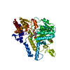
| ||||||||
|---|---|---|---|---|---|---|---|---|---|
| 1 | 
| ||||||||
| Unit cell |
|
-Components
| #1: Protein | Mass: 46503.246 Da / Num. of mol.: 1 Source method: isolated from a genetically manipulated source Source: (gene. exp.) Staphylococcus aureus (bacteria) / Gene: sufS / Production host: | ||||
|---|---|---|---|---|---|
| #2: Chemical |   Mass: 62.068 Da / Num. of mol.: 3 / Source method: obtained synthetically / Formula: C2H6O2 Mass: 62.068 Da / Num. of mol.: 3 / Source method: obtained synthetically / Formula: C2H6O2#3: Water | ChemComp-HOH / |  Mass: 18.015 Da / Num. of mol.: 369 / Source method: isolated from a natural source / Formula: H2O Mass: 18.015 Da / Num. of mol.: 369 / Source method: isolated from a natural source / Formula: H2OHas ligand of interest | Y | |
-Experimental details
-Experiment
| Experiment | Method: X-RAY DIFFRACTION / Number of used crystals: 1 |
|---|
- Sample preparation
| Crystal | Density Matthews: 2.77 Å3/Da / Density % sol: 55.66 % |
|---|---|
| Crystal grow | Temperature: 293 K / Method: vapor diffusion, sitting drop / pH: 8.35 / Details: Tris, ammonium sulfate |
-Data collection
| Diffraction | Mean temperature: 100 K / Serial crystal experiment: N |
|---|---|
| Diffraction source | Source: SEALED TUBE / Type: BRUKER D8 QUEST / Wavelength: 1.34 Å |
| Detector | Type: Bruker PHOTON III / Detector: PIXEL / Date: Nov 16, 2021 / Details: Helios MX |
| Radiation | Protocol: SINGLE WAVELENGTH / Monochromatic (M) / Laue (L): M / Scattering type: x-ray |
| Radiation wavelength | Wavelength: 1.34 Å / Relative weight: 1 |
| Reflection | Resolution: 1.388→31.737 Å / Num. obs: 104403 / % possible obs: 100 % / Redundancy: 22.6 % / Rmerge(I) obs: 0.015 / Rpim(I) all: 0.015 / Rrim(I) all: 0.021 / Net I/σ(I): 22.8 |
| Reflection shell | Resolution: 1.388→1.41 Å / Redundancy: 1.9 % / Rmerge(I) obs: 0.415 / Mean I/σ(I) obs: 2.1 / Num. unique obs: 4995 / Rpim(I) all: 0.415 / Rrim(I) all: 0.587 / % possible all: 97.6 |
- Processing
| Software |
| |||||||||||||||||||||||||||||||||||||||||||||||||||||||||||||||||||||||||||||||||||||||||||||||||||||||||||||||||||||||||||||||||||||||||||||||||||||||||||
|---|---|---|---|---|---|---|---|---|---|---|---|---|---|---|---|---|---|---|---|---|---|---|---|---|---|---|---|---|---|---|---|---|---|---|---|---|---|---|---|---|---|---|---|---|---|---|---|---|---|---|---|---|---|---|---|---|---|---|---|---|---|---|---|---|---|---|---|---|---|---|---|---|---|---|---|---|---|---|---|---|---|---|---|---|---|---|---|---|---|---|---|---|---|---|---|---|---|---|---|---|---|---|---|---|---|---|---|---|---|---|---|---|---|---|---|---|---|---|---|---|---|---|---|---|---|---|---|---|---|---|---|---|---|---|---|---|---|---|---|---|---|---|---|---|---|---|---|---|---|---|---|---|---|---|---|---|
| Refinement | Method to determine structure: MOLECULAR REPLACEMENT Starting model: 5J8Q Resolution: 1.388→31.737 Å / Cor.coef. Fo:Fc: 0.973 / Cor.coef. Fo:Fc free: 0.966 / SU B: 0.863 / SU ML: 0.034 / Cross valid method: FREE R-VALUE / ESU R: 0.049 / ESU R Free: 0.051 Details: Hydrogens have been added in their riding positions
| |||||||||||||||||||||||||||||||||||||||||||||||||||||||||||||||||||||||||||||||||||||||||||||||||||||||||||||||||||||||||||||||||||||||||||||||||||||||||||
| Solvent computation | Ion probe radii: 0.8 Å / Shrinkage radii: 0.8 Å / VDW probe radii: 1.2 Å / Solvent model: MASK BULK SOLVENT | |||||||||||||||||||||||||||||||||||||||||||||||||||||||||||||||||||||||||||||||||||||||||||||||||||||||||||||||||||||||||||||||||||||||||||||||||||||||||||
| Displacement parameters | Biso mean: 17.802 Å2
| |||||||||||||||||||||||||||||||||||||||||||||||||||||||||||||||||||||||||||||||||||||||||||||||||||||||||||||||||||||||||||||||||||||||||||||||||||||||||||
| Refinement step | Cycle: LAST / Resolution: 1.388→31.737 Å
| |||||||||||||||||||||||||||||||||||||||||||||||||||||||||||||||||||||||||||||||||||||||||||||||||||||||||||||||||||||||||||||||||||||||||||||||||||||||||||
| Refine LS restraints |
| |||||||||||||||||||||||||||||||||||||||||||||||||||||||||||||||||||||||||||||||||||||||||||||||||||||||||||||||||||||||||||||||||||||||||||||||||||||||||||
| LS refinement shell |
|
