 Movie
Movie Controller

[English] 日本語
 Yorodumi
- PDB-8d7p: Human Casein kinase 1 delta in complex with phosphorylated Drosop... -
+ Open data

- Basic information
| Entry | Database: PDB / ID: 8d7p | ||||||
|---|---|---|---|---|---|---|---|
| Title | Human Casein kinase 1 delta in complex with phosphorylated Drosophila PERIOD peptide | ||||||
 Components Components |
| ||||||
 Keywords Keywords | TRANSFERASE/CIRCADIAN CLOCK PROTEIN / Transferase / CIRCADIAN CLOCK PROTEIN / Kinase / complex / TRANSFERASE-CIRCADIAN CLOCK PROTEIN complex | ||||||
| Function / homology |  Function and homology information Function and homology informationpositive regulation of non-canonical Wnt signaling pathway / protein localization to Golgi apparatus / COPII vesicle coating / microtubule nucleation / midbrain dopaminergic neuron differentiation / tau-protein kinase / protein localization to cilium / non-motile cilium assembly / protein localization to centrosome / COPII-mediated vesicle transport ...positive regulation of non-canonical Wnt signaling pathway / protein localization to Golgi apparatus / COPII vesicle coating / microtubule nucleation / midbrain dopaminergic neuron differentiation / tau-protein kinase / protein localization to cilium / non-motile cilium assembly / protein localization to centrosome / COPII-mediated vesicle transport / tau-protein kinase activity / Golgi organization / Major pathway of rRNA processing in the nucleolus and cytosol / : / spindle assembly / endoplasmic reticulum-Golgi intermediate compartment membrane / Loss of Nlp from mitotic centrosomes / Loss of proteins required for interphase microtubule organization from the centrosome / Recruitment of mitotic centrosome proteins and complexes / Recruitment of NuMA to mitotic centrosomes / Anchoring of the basal body to the plasma membrane / AURKA Activation by TPX2 / spindle microtubule / circadian regulation of gene expression / regulation of circadian rhythm / spindle / endocytosis / Wnt signaling pathway / Regulation of PLK1 Activity at G2/M Transition / positive regulation of canonical Wnt signaling pathway / positive regulation of proteasomal ubiquitin-dependent protein catabolic process / actin cytoskeleton / protein phosphorylation / protein kinase activity / non-specific serine/threonine protein kinase / cilium / ciliary basal body / cadherin binding / protein serine kinase activity / protein serine/threonine kinase activity / centrosome / perinuclear region of cytoplasm / Golgi apparatus / signal transduction / nucleoplasm / ATP binding / nucleus / plasma membrane / cytoplasm / cytosol Similarity search - Function | ||||||
| Biological species |  Homo sapiens (human) Homo sapiens (human) | ||||||
| Method |  X-RAY DIFFRACTION / SYNCHROTRON / MOLECULAR REPLACEMENT / Resolution: 2.25 Å X-RAY DIFFRACTION / SYNCHROTRON / MOLECULAR REPLACEMENT / Resolution: 2.25 Å | ||||||
 Authors Authors | Philpott, J.M. / Freeberg, A.M. / Tripathi, S.M. / Partch, C.L. | ||||||
| Funding support |  United States, 1items United States, 1items
| ||||||
 Citation Citation | Journal: Mol.Cell / Year: 2023 Title: PERIOD phosphorylation leads to feedback inhibition of CK1 activity to control circadian period. Authors: Philpott, J.M. / Freeberg, A.M. / Park, J. / Lee, K. / Ricci, C.G. / Hunt, S.R. / Narasimamurthy, R. / Segal, D.H. / Robles, R. / Cai, Y. / Tripathi, S. / McCammon, J.A. / Virshup, D.M. / ...Authors: Philpott, J.M. / Freeberg, A.M. / Park, J. / Lee, K. / Ricci, C.G. / Hunt, S.R. / Narasimamurthy, R. / Segal, D.H. / Robles, R. / Cai, Y. / Tripathi, S. / McCammon, J.A. / Virshup, D.M. / Chiu, J.C. / Lee, C. / Partch, C.L. | ||||||
| History |
|
- Structure visualization
| Structure viewer | Molecule: MolmilJmol/JSmol |
|---|
- Downloads & links
-Download
| PDBx/mmCIF format | 8d7p.cif.gz | 135.5 KB | Display | PDBx/mmCIF format |
|---|---|---|---|---|
| PDB format | pdb8d7p.ent.gz | 103.6 KB | Display | PDB format |
| PDBx/mmJSON format | 8d7p.json.gz | Tree view | PDBx/mmJSON format | |
| Others |  Other downloads Other downloads |
-Validation report
| Arichive directory | https://data.pdbj.org/pub/pdb/validation_reports/d7/8d7pftp://data.pdbj.org/pub/pdb/validation_reports/d7/8d7p | HTTPS FTP |
|---|
-Related structure data
| Related structure data | 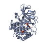 8d7mC 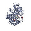 8d7nC 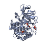 8d7oC 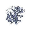 6pxoS S: Starting model for refinement C: citing same article ( |
|---|---|
| Similar structure data |
-Links
 PDBj
PDBj


- Assembly
| Deposited unit | 
| ||||||||
|---|---|---|---|---|---|---|---|---|---|
| 1 | 
| ||||||||
| 2 | 
| ||||||||
| Unit cell |
|
-Components
| #1: Protein | Mass: 34954.344 Da / Num. of mol.: 2 Source method: isolated from a genetically manipulated source Source: (gene. exp.) Homo sapiens (human) / Gene: CSNK1D, HCKID / Production host:  References: UniProt: P48730, non-specific serine/threonine protein kinase, tau-protein kinase #2: Protein/peptide | Mass: 2122.232 Da / Num. of mol.: 2 / Source method: obtained synthetically / Source: (synth.) #3: Water | ChemComp-HOH / |  Mass: 18.015 Da / Num. of mol.: 135 / Source method: isolated from a natural source / Formula: H2O Mass: 18.015 Da / Num. of mol.: 135 / Source method: isolated from a natural source / Formula: H2OHas ligand of interest | Y | Has protein modification | Y | |
|---|
-Experimental details
-Experiment
| Experiment | Method: X-RAY DIFFRACTION / Number of used crystals: 1 |
|---|
- Sample preparation
| Crystal | Density Matthews: 2.34 Å3/Da / Density % sol: 42.75 % |
|---|---|
| Crystal grow | Temperature: 295 K / Method: vapor diffusion, hanging drop / pH: 5.5 / Details: 0.16M Succinic Acid (5.5) 23% PEG3350 |
-Data collection
| Diffraction | Mean temperature: 100 K / Serial crystal experiment: N |
|---|---|
| Diffraction source | Source: SYNCHROTRON / Site: APS / Beamline: 23-ID-D / Wavelength: 1.03 Å |
| Detector | Type: DECTRIS PILATUS 6M / Detector: PIXEL / Date: Dec 10, 2021 |
| Radiation | Protocol: SINGLE WAVELENGTH / Monochromatic (M) / Laue (L): M / Scattering type: x-ray |
| Radiation wavelength | Wavelength: 1.03 Å / Relative weight: 1 |
| Reflection | Resolution: 2.25→60.77 Å / Num. obs: 27457 / % possible obs: 93.7 % / Redundancy: 3.1 % / CC1/2: 0.98 / Rmerge(I) obs: 0.116 / Net I/σ(I): 5.5 |
| Reflection shell | Resolution: 2.25→2.32 Å / Rmerge(I) obs: 0.312 / Mean I/σ(I) obs: 2.7 / Num. unique obs: 2505 / CC1/2: 0.85 |
- Processing
| Software |
| ||||||||||||||||||||||||||||||||||||||||||||||||||||||||||||||||||||||
|---|---|---|---|---|---|---|---|---|---|---|---|---|---|---|---|---|---|---|---|---|---|---|---|---|---|---|---|---|---|---|---|---|---|---|---|---|---|---|---|---|---|---|---|---|---|---|---|---|---|---|---|---|---|---|---|---|---|---|---|---|---|---|---|---|---|---|---|---|---|---|---|
| Refinement | Method to determine structure: MOLECULAR REPLACEMENT Starting model: 6PXO Resolution: 2.25→48.514 Å / SU ML: 0.27 / Cross valid method: FREE R-VALUE / σ(F): 1.97 / Phase error: 28.66 / Stereochemistry target values: ML
| ||||||||||||||||||||||||||||||||||||||||||||||||||||||||||||||||||||||
| Solvent computation | Shrinkage radii: 0.9 Å / VDW probe radii: 1.11 Å / Solvent model: FLAT BULK SOLVENT MODEL | ||||||||||||||||||||||||||||||||||||||||||||||||||||||||||||||||||||||
| Refinement step | Cycle: LAST / Resolution: 2.25→48.514 Å
| ||||||||||||||||||||||||||||||||||||||||||||||||||||||||||||||||||||||
| Refine LS restraints |
| ||||||||||||||||||||||||||||||||||||||||||||||||||||||||||||||||||||||
| LS refinement shell |
|
